
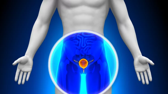
For prostate cancer diagnostics, 7T MRI has next to nothing on ultrasound tomography
In an initial comparison study involving 10 patients with high-risk prostate cancer, ultrasound tomography (UT) soundly beat 7-Tesla multiparametric MRI on detection sensitivity, 85.7% to 65.3%.
While the experimental modality trailed the MRI on specificity in the study, it wasn’t far behind, 93.9% vs. 96.6%.
The study was conducted by researchers from the National Cancer Institute and Johns Hopkins University and is being publicized by the manufacturer of the UT technology used in the comparison, QT Imaging Inc.
In an abstract recognized as a “best of 2022” by the Engineering and Urology Society, lead author Jacob Enders, a medical student researcher with the NCI’s urologic oncology branch, senior author Peter Pinto, MD, an investigator and fellowship director in the branch, and colleagues place the project in context.
They write: “While multiparametric MRI (mpMRI) and targeted biopsy have improved the detection of high-risk prostate cancer (PCa) over traditional B-mode ultrasound, the use of mpMRI in the diagnostic pathway for PCa remains limited due to variability in image acquisition and interpretation, accessibility and cost” [1].
The authors cite previous research demonstrating quantitative transmission UT’s high sensitivity for diagnosing breast cancer.
The burgeoning technology, they note, offers “360-degree submillimeter-resolution imaging for tissue characterization and cancer detection, even in the presence of bone and air, or with limited view tomographic data.”
For the study, the researchers used ex vivo prostate glands of men who had undergone radical prostatectomy.
Comparing lesion detection and visualization between UT and MRI, along with histopathology annotations, was the primary point of the exercise.
Images were acquired within 30 minutes of surgery completion, and tumor boundaries were segmented on both modalities by an expert genitourinary radiologist.
Meanwhile two expert genitourinary pathologists created wholemount pathology slides with patient-specific molds.
The researchers used an 18-sector approach that divided the prostate into anatomical regions to correlate pathology areas of interest with those in imaging.
They defined sensitivity as “the probability of correctly detecting and identifying tumor-positive regions on a given modality (UT or mpMRI) compared with ground truth histopathology.”
Specificity was defined as “the probability of correctly identifying tumor-negative regions.”
In a news release, study co-author John Klock, MD, the CEO and CMO of QT Imaging, says the results “demonstrate the clinical value of QT’s low frequency transmitted sound volography as a high-resolution imaging modality.”

Dave P. has worked in journalism, marketing and public relations for more than 30 years, frequently concentrating on hospitals, healthcare technology and Catholic communications. He has also specialized in fundraising communications, ghostwriting for CEOs of local, national and global charities, nonprofits and foundations.