Computed Tomography

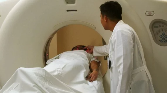
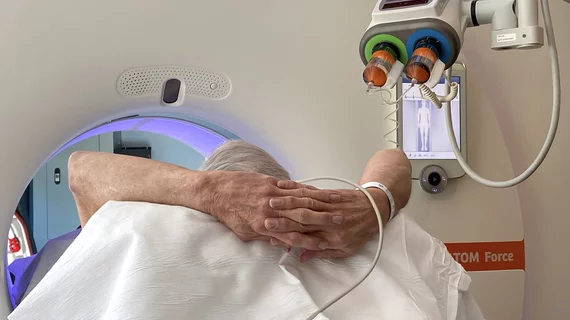
Computed tomography (CT) is a fast and accurate imaging modality often used in emergency settings and trauma imaging. CT scans, with or without (or both) iodinated contrast are frequently used to image the brain, chest, abdomen and pelvis, but also have post-imaging reconstructive capabilities for detailed orthopedic imaging. It is now a standard imaging modality in emergency rooms to quickly assess patients. CT uses a series of X-ray images shot as the gantry rotates around the patient. Computer technology assembles these into into a dataset volume than can be slices on any access, or advanced visualization software can extract specific parts of the anatomy for study. Find more content specific to cardiac CT.

Vital Images Highlighting latest version of Vitrea Advanced Visualization at SCCT Scientific Meeting

MINNETONKA, Minn., June 29, 2017 (GLOBE NEWSWIRE) -- Vital Images is highlighting the benefits of its Vitrea solutions as it participates in the 12th Annual Scientific Meeting of the Society of Cardiovascular Computed Tomography, July 6-9, in Washington, D.C., at booth 401.

Society of Cardiovascular Computed Tomography (SCCT) Announces New Fellows and Residents-In-Training Partnership with Toshiba Medical

The Society of Cardiovascular Computed Tomography (SCCT) and Toshiba Medical today announce a new partnership dedicated to providing career development opportunities for radiology fellows and residents in the United States. Toshiba Medical’s support will allow 250 U.S.-based fellows and residents to join SCCT at no charge for one year, enhancing their early career opportunities and building their awareness of the growing contributions of cardiovascular computed tomography.

Intrinsic Imaging awarded interventional medical device trial for prevention of pulmonary emboli

Intrinsic Imaging, an FDA audited, ISO 9001:2008 and ISO 13485:2003 certified, GAMP 5 compliant medical imaging core lab specializing in providing services for clinical trials, announced today that it has been awarded a clinical trial to study an interventional medical device designed for the prevention of pulmonary emboli.

Example of a curved MPR image reconstruction of entire length of of a coronary artery on a cardiac CT scan to better show calcified and soft plaque burden inside the vessel. The thumbnail dots on the left side of the image are cross sectional views of the vessel. Siemens example on the expo floor.

The CCTA Playbook: A Guide to Coding, Reimbursement and Operations

A compendium of the business intelligence required to launch a CCTA service

Be Mindful of the Stark Law in CCTA Alliances

Numerous radiology groups are currently discussing arrangements whereby cardiologists and the radiologists would essentially split the responsibilities for reading coronary CT angiography (CCTA) studies. Most commonly, as proposed, the cardiologists would read and generate a signed interpretation report, but limited to the cardiac portion of the

Around the web

The nuclear imaging isotope shortage of molybdenum-99 may be over now that the sidelined reactor is restarting. ASNC's president says PET and new SPECT technologies helped cardiac imaging labs better weather the storm.

CMS has more than doubled the CCTA payment rate from $175 to $357.13. The move, expected to have a significant impact on the utilization of cardiac CT, received immediate praise from imaging specialists.

The all-in-one Omni Legend PET/CT scanner is now being manufactured in a new production facility in Waukesha, Wisconsin.

Trimed Popup
Trimed Popup