
Bacteria-based contrast agent offers 'substantial improvements' for tumor border delineation during surgery
Experts have developed a bacteria-based contrast agent they believe can significantly enhance the surgical removal of tumors.
The agent is the result of a collaboration between experts at the Korea Institute of Science and Technology (KIST) and Chungnam National University Hospital, in South Korea. It was developed with surgeons in mind, as completely removing tumors and their margins represents a significant challenge, and the failure to do so often results in additional surgeries.
“Current imaging techniques suffer from a lack of specificity and resolution, leading to inaccurate tumor imaging and limited applicability of targeted contrast agents, as they require cancer-specific development,” SeungBeum Suh, PhD, from the Center for Bionics at KIST, and colleagues explained in Advanced Materials. “The need for enhanced contrast through improved tumor-to-background ratio (TBR) and the toxicity from repeated injections due to fading fluorescent signals further complicate the issue. Additionally, challenges in visualizing the entire 3D tumor with surface-stained contrast agents highlight the demand for advanced imaging solutions for more precise surgical guidance.”
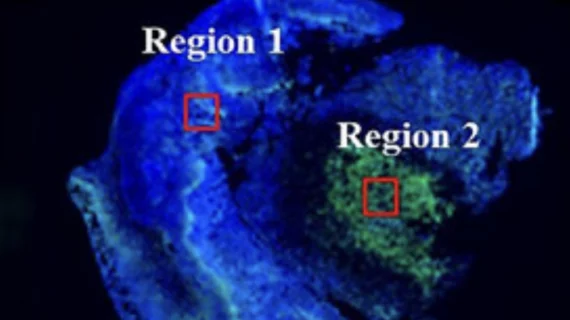
The fluorescent agent is made from Streptavidin Associated Salmonella (SAS), which can attach to cancerous tissues and secrete streptavidin upon induction; when combined with biotin-conjugated fluorescent dyes (streptavidin binds strongly to biotin) and injected into a tumor, the bacteria will light up the area—margins included—surgeons are seeking to remove. Essentially, it places a bright neon sign on the entirety of the tumor and its borders, and it does so autonomously.
There are two key benefits the bacteria-based agent has over other contrast agents: longevity and luminescence. After injection, it remains visible to the naked eye for up to 72 hours, and it is said to be five times brighter than other agents used for the same purpose. What’s more, the system operates in the near-infrared spectrum, which makes it compatible with standard endoscopic and imaging equipment.
So far, the agent has proven to be both safe and effective, while also displaying stable physiological responses. Researchers are hopeful it can be safely implemented into clinical settings beyond just tumor delineation during surgical procedures, but potentially in precision drug delivery as well.
“This innovative method offers substantial improvements over existing fluorescent contrast agents and holds promise for both diagnostic and therapeutic applications in cancer surgery,” the group concluded.
Read more about the innovative imaging method here.

In addition to her background in journalism, Hannah also has patient-facing experience in clinical settings, having spent more than 12 years working as a registered rad tech. She began covering the medical imaging industry for Innovate Healthcare in 2021.