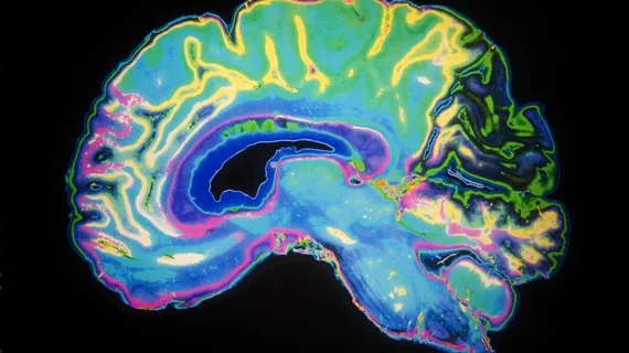
Depression treatments can change the brain’s infrastructure in just 6 weeks
New research involving MR images acquired before and after depression treatments has revealed that adult patients with major depressive disorder experience changes in the structural connectivity of their brains after undergoing just six weeks of inpatient treatments.
Moreover, patients who respond well to therapy show greater increases in brain connectivity than those who do not.
The surprising findings, presented by Jonathan Repple, MD, at the 35th European College of Neuropsychopharmacology (ECNP) in Vienna, indicate that the adult brain may be much less rigid or “fixed” than many scientists previously thought.
“We found that treatment for depression changed the infrastructure of the brain, which goes against previous expectations,” says Repple, a professor of predictive psychiatry at the University of Frankfurt, in a news release promoting the research. “This gives hope to patients who believe nothing can change and they have to live with a disease forever, because it is ‘set in stone’ in their brain.”
The 105 patients with depression in the study received different treatments: electroconvulsive therapy, psychological therapy, medication or a combination of all therapies. The results showed increased brain connectivity to be consistent across the various therapies and undifferentiated by therapy types.
We don’t have an explanation as to how these changes take place, or why they should happen with such different forms of treatment,” Repple says.
The study included a group of 55 healthy controls. The researchers found this cohort did not see significant changes in connectivity over time.
“A second scan showing that there are no time effects in healthy controls supports our findings that we see something that is related to the disease and more importantly the treatment of this disease,” Repple said.
While the research provides interesting insights into the brain’s flexibility, it also raises questions. Psychiatrist Eric Ruhe, MD, PhD, of Rabdoud University Medical Center in the Netherlands calls for clinical inquiry into whether “different treatments have the possibility to specifically change targeted brain networks or, vice versa, whether we can use the disturbances in brain networks as measured in the present study to choose which therapy will be helpful.”
The full news release is available for downloading here.
