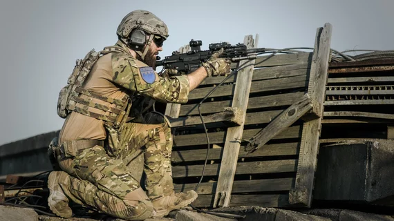
Functional MRI shows war veterans’ brains compensating for blast-related TBI
The brains of war veterans who have suffered blast-related mild traumatic brain injury (mTBI) appear to ward off long-term memory loss by changing connectivity across multiple regions, according to a pilot study published online Dec. 5 in Brain Imaging and Behavior.
Lead author Kathleen Pagulayan, PhD, of the VA Northwest Health Network and the University of Washington in Seattle and colleagues noted the commonality of working-memory loss among veterans who have survived explosions and the difficulty of quantifying the associated mTBI using neuropsychological examinations.
The team sought to improve understanding of the problem by adding functional MRI, which would allow visual comparison of the resting-state working memory systems of 24 previously deployed blast survivors who have mTBI and 17 veterans with no history of brain injury.
The team first evaluated all subjects’ working memory with a Auditory Consonant Trigrams (ACT) test. The subjects were then imaged with resting-state fMRI. The researchers homed in on functional connectivity from parts of the brain known as frontal “seed regions,” which are known components of the working memory network.
Pagulayan and co-authors found no significant group differences on the ACT test. However, they reported, neuroradiologist interpreters of the fMRI scans found significant hyper-connectivity from frontal seed regions in the veterans with a history of mTBI. Such heightened connectivity was not present in the control group.
What’s more, within the mTBI group alone, better ACT performance correlated with increased functional connectivity to multiple brain regions. These included cerebellar components of the working memory network.
The authors noted that these results were present after controlling for age, PTSD symptoms and estimated premorbid IQ.
Long-term alterations in the functional connectivity of the working memory network following blast-related mTBI, Pagulayan et al suggested, “may reflect a compensatory change that contributes to intact performance on an objective measure of working memory.”

