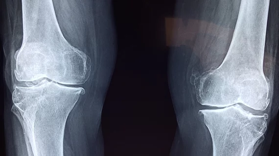
AI detects early signs of osteoarthritis from X-ray images
By examining X-ray images for tibial spiking in the knee joint, an AI-based neural network was able to detect early knee osteoarthritis that overlapped with 87% of doctors’ diagnoses.
The findings, published in a new study in Diagnostics, offer a way to potentially improve early detection of osteoarthritis, which in turn may allow patients to take measures to prevent the condition from getting worse. Although tibial spiking is not currently included in official diagnostic criteria for osteoarthritis, the study notes that orthopedic specialists typically consider it a predictive radiological feature.
“The aim of the project was to train the AI to recognize an early feature of osteoarthritis from an x-ray. Something that experienced doctors can visually distinguish from the image, but cannot be done automatically,” explains Anri Patron, the researcher responsible for the development of the method, in a news release. “Around 700 x-ray images were used in developing the AI model, after which the model was validated with around 200 x-ray images.”
Overall, says Sami Äyrämö, head of the Digital Health Intelligence Laboratory at the University of Jyväskylä in Finland, the research and development of the new AI model helps fill in existing gaps in AI-based detection, since previously existing models had been more focused on later-stage diagnoses.
“Several AI models have previously been developed to detect knee osteoarthritis. These models can detect severe cases that would be easily detected by any specialists. However the previously developed methods are not accurate enough to detect the early-stage manifestations. The method now being developed aims for, in particular, early detection from x-rays, for which there is a great need,” Äyrämö says.
Early diagnosis of osteoarthritis not only offers medical benefits for patients—such as giving patients a chance to treat symptoms and slow down the progression of the disease—but it also may help reduce the necessity for more expensive examinations, such as MRI.
“In the best possible scenario, the patient might even avoid joint replacement surgery,” says Central Finland Health Care District CEO and professor of surgery Juha Paloneva.
