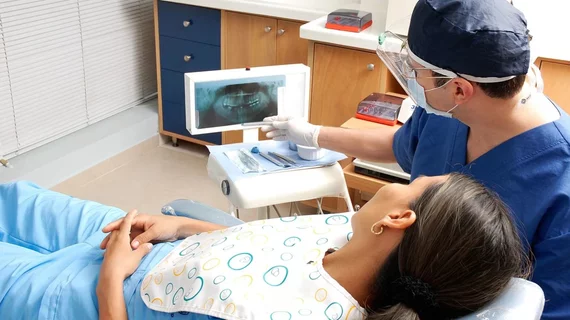
Dental x-rays may be causing hundreds of excess cancer cases each year
Dental x-ray exams may be causing hundreds of excess cancer cases in the U.S. each year, according to a new analysis.
Researchers pinpoint the number at 967 instances of the disease, occurring in the head and neck regions. Most such cancers stem from full-mouth x-ray series and dental cone-beam CT scans, noted experts with Creighton and Ohio state universities.
That number could be cut by 75% (down to 237) through rectangular collimation to limit the x-ray beam’s size and stricter selection criteria to weed out unnecessary scans.
“There should never be prescription of radiographs before a clinical examination,” first author Douglas Benn, DDS, PhD, a radiologist and professor of oral and maxillofacial imaging at Creighton University in Omaha, Nebraska, advised. “Unfortunately, it is well-documented that most dentists will image patients on a routine (annual or semi-annual) basis, including those patients at low for dental caries,” he added later.
To reach their conclusions, Benn and co-author Peter Vig analyzed totals derived from the 2014-2015 Nationwide Evaluation of X-Ray Trends survey of dental offices. They also incorporated data from the Medical Radiation Exposure of Patients in the U.S. report and other sources. The authors estimated that only 1% of general dental offices use collimators or paperwork gaining patients’ approval for radiological procedures.
“Use of an informed consent form containing sufficient information to help the patient and dentist understand the risk of cancer formation may help to reduce the over prescription of dental radiographs,” Benn and Vig wrote.
You can read more about the analysis here in the journal of Oral Surgery, Oral Medicine, Oral Pathology and Oral Radiology.

