Medical Imaging

Physicians utilize medical imaging to see inside the body to diagnose and treat patients. This includes computed tomography (CT), magnetic resonance imaging (MRI), X-ray, ultrasound, fluoroscopy, angiography,  and the nuclear imaging modalities of PET and SPECT. 

Thumbnail

These deep learning algorithms outperformed a panel of 11 pathologists

During a 2016 simulation exercise, researchers evaluated the ability of 32 different deep learning algorithms to detect lymph node metastases in patients with breast cancer. Each algorithm’s performance was then compared to that of a panel of 11 pathologists with time constraint (WTC). Overall, the team found that seven of the algorithms outperformed the panel of pathologists, publishing an in-depth analysis in JAMA.

Radiology professor uses nanotechnology to develop potential cancer treatment

Kattesh V. Katti, MScEd, PhD, a professor of radiology and physics at the University of Missouri in Columbia, specializes in planet-friendly nanotechnology and has developed a potential cancer treatment that “uses no toxic chemicals and leaves zero toxic waste.”

Radiologists scan mummy to better understand the past

A 2,000-year-old Egyptian mummy named Hen, from the Cazenovia Public Library in New York, was transferred for five hours to the Crouse Hinds Hospital in Syracuse for medical tests and technological exams that could potentially offer new information about the mummy and humanity in that time.

RSNA 2017: Are EDs utilizing breast ultrasound too often?

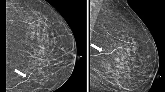
An abscess is one of the few breast-specific emergencies that require urgent treatment in emergency departments (EDs). A new study, presented at RSNA 2017 in Chicago, found that ED environments generate unnecessary exams in addition to some that incompletely identify malignancies.

Patient shares struggles with being claustrophobic and undergoing MRI

It’s a problem radiologists and technologists encounter from time to time: the claustrophobic patient who needs an MRI. They need the exam, but they’re not exactly happy about the process.

Novel needle technique takes optical-ultrasound guidance inside the heart

British researchers have successfully outfitted a needle with optical fibers capable of transmitting and receiving ultrasonic pulses within the heart during minimally invasive cardiac surgeries. The achievement may represent a new way to optically image tiny tissue targets in real time and at high resolution throughout the body.

UK’s NHS radiology services under review after 20,000 X-rays go unread

The National Health Service’s Care Quality Commission has launched a national review of its radiology services after indications that more than 20,000 chest x-rays and 2,000 abdomen X-rays of patients were not interpreted by trained personnel.

Thumbnail

RSNA 2017: Overuse of STAT designation slows MRI workflow, causes confusion

The term “STAT” implies urgency: act immediately. But when clinicians overuse that designation, it becomes more difficult and time-consuming to distinguish true emergencies from cases that can wait, said Anna Trofimova, MD, PhD, a diagnostic radiology resident at Emory University School of Medicine.

Around the web

The patient, who was being cared for in the ICU, was not accompanied or monitored by nursing staff during his exam, despite being sedated.

The nuclear imaging isotope shortage of molybdenum-99 may be over now that the sidelined reactor is restarting. ASNC's president says PET and new SPECT technologies helped cardiac imaging labs better weather the storm.

CMS has more than doubled the CCTA payment rate from $175 to $357.13. The move, expected to have a significant impact on the utilization of cardiac CT, received immediate praise from imaging specialists.