Medical Imaging

Physicians utilize medical imaging to see inside the body to diagnose and treat patients. This includes computed tomography (CT), magnetic resonance imaging (MRI), X-ray, ultrasound, fluoroscopy, angiography,  and the nuclear imaging modalities of PET and SPECT. 

Dosimetry using average-sized phantoms leads to considerable errors

The use of non-size-dependent reference phantoms to calculate CT dose can lead to errors in calculating the radiation received by a patient, according to an article published in the American Journal of Roentgenology. Using data from the National Lung Screening Trial, researchers found using an average height and weight to calculate dose can lead to errors of up to 200 percent when compared with a more accurate estimate using a particular patient’s height and weight. 

X-ray scanners in New Hampshire prison may pose health concerns amongst other things

X-ray machines in New Hampshire’s prisons were initially brought in to stop the influx of drugs. But a year later, these machines await installation due to health concerns and the language used in the bill that passed last year.

Carestream Demonstrates Touch Prime Ultrasound Systems at American Institute of Ultrasound in Medicine Convention

System Design Enhances Views of Moving Structures, Eliminates Angle Limitations of Traditional Doppler Ultrasound

Researchers working to develop MRI-powered mini robots to deliver treatment

Researchers from the University of Houston and Houston Methodist Hospital are working to develop and deploy MRI-powered mini robots to travel the body’s venous system delivering drugs or a self-assembled interventional tool as treatment.

Thumbnail

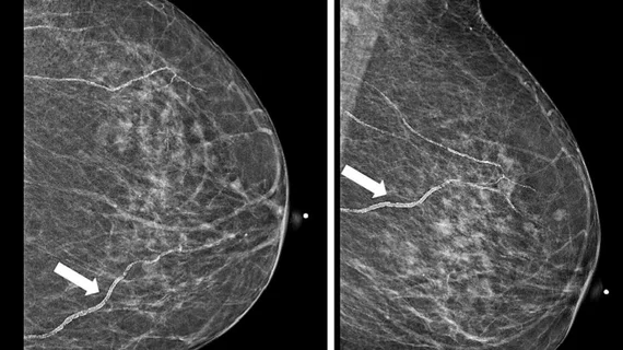
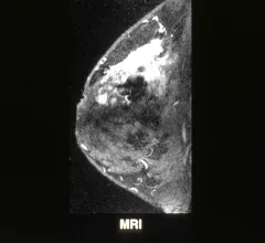
MRI useful for second look at inconclusive mammograms

MRI can be a useful tool for taking a second look at mammograms that are considered inconclusive in a process known as “problem-solving MRI," but radiologists should be cautious about the possibility of false negatives, according to a study published in the American Journal of Roentgenology.

Toshiba Medical’s Newest MRI System Tapped for UCI Brain Trauma Research

TUSTIN, Calif., March 8, 2017 – Researchers at the University of California, Irvine now have access to Toshiba Medical’s most advanced MR technology for their cutting edge neurology work investigating cerebral microbleeds (CMB) in high school football players as a potential precursor to chronic traumatic encephalopathy (CTE). As part of its ongoing, first-of-its-kind study to identify early signs of brain trauma in young athletes, UCI doctors will now leverage Toshiba Medical’s all-new Vantage GalanTM 3T MR system as a key diagnostic tool.

Qioptiq Introduces SlimLine Camera Lens System for Mobile X-Ray C-Arms

Turnkey Sub-Assembly is Cost-Optimized for Streamlined Integration and High Quality Imaging at Value-Added Price Point

Siemens Healthineers Paves the Way for Precision Medicine With BioMatrix Technology and Magnetom Vida MRI Scanner

New BioMatrix technology adapts automatically to individual anatomical and physiological characteristics to offer consistent, high-quality imaging for all patients

Around the web

The ACR hopes these changes, including the addition of diagnostic performance feedback, will help reduce the number of patients with incidental nodules lost to follow-up each year.

And it can do so with almost 100% accuracy as a first reader, according to a new large-scale analysis.

The patient, who was being cared for in the ICU, was not accompanied or monitored by nursing staff during his exam, despite being sedated.