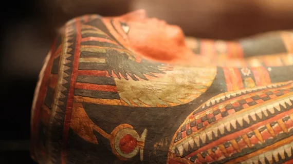
Researchers use CT scans to develop portraits of ancient Egyptians
With the help of current CT technology, along with computer graphics and craniofacial reconstruction, a team of Johns Hopkins researchers were able to create detailed portraits of the mummified remains of an ancient Egyptian, according to reporting from The Baltimore Sun.
Elliot K. Fishman, MD, a professor of radiology and the director of diagnostic imaging at Johns Hopkins Hospital, helped generate CT scans of the mummified remains.
The CT scans were eventually sent to a forensic facial reconstruction laboratory and were developed into portraits, which are now part of the “Who Am I? Remembering The Dead Through Facial Reconstruction” exhibition at Baltimore's Johns Hopkins Archaeological Museum.
“These women look at you the moment you walk in the door; you’re looking at them, and they’re looking at you,” said Sanchita Balachandran, the museum’s associate director. “It feels as though they’re right here with us.”
To read the story on The Baltimore Sun’s website, click the link below.

