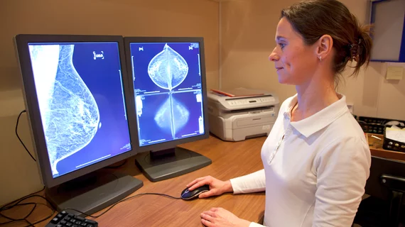
How does CAD-enhanced synthetic mammography compare to FFDM?
Combining digital breast tomosynthesis (DBT) and standard 2D full-field digital mammography (FFDM) gives radiologists a powerful tool for detecting deadly breast lesions. A downside of this combination, however, is that it means exposing the patient to much more radiation.
Two possible alternatives to using both DBT and FFDM are generating a synthetic 2D mammogram from the DBT data set or creating a computer-aided detection (CAD)-enhanced synthetic mammogram—but can either alternative really replace FFDM?
A new study from Clinical Radiology compared the diagnostic performance of CAD-enhanced synthetic mammography, synthetic mammography and FFDM to see which modality produced the best 2D images. The authors gathered data from 68 cases where all three mammogram types were available, asking two experienced radiologists—blinded to image type and final assessment outcome—to retrospectively read the exams and assign a score from one to five for each pair of images. Out of the 68 cases, 34 were malignancies and the other 34 were normal or benign.
Overall, the diagnostic accuracy of the CAD-enhanced synthetic mammogram was “significantly improved” compared to the standard synthetic mammogram and FFDM mammogram. The area under the ROC curve was 0.846 for CAD-enhanced synthetic mammography, 0.724 for FFDM and 0.683 for standard synthetic mammography.
The CAD-enhanced synthetic mammogram also had the highest diagnostic accuracy for “masses, parenchymal deformities and asymmetric densities.” In addition, the CAD-enhanced synthetic mammogram and FFDM both performed “significantly better” than the standard synthetic mammogram when it came to assessing mass lesions.
“These results show that CAD enhancement can improve the diagnostic performance of the synthetic mammogram, potentially delivering performance that is superior to the conventional 2D FFDM,” wrote lead author J.J. James, with the Nottingham Breast Institute in Nottingham, England, and colleagues. “This is achieved through improved specificity compared to 2D FFDM and improved sensitivity compared to the standard synthetic mammogram.”
More research is needed to determine how the CAD-enhanced synthetic images could be worked into the DBT screening workflow, the authors added.
“Although the performance of the CAD-enhanced synthetic mammogram was superior to the standard synthetic image, it is unclear as to whether the CAD-enhanced synthetic image is best used as a replacement for the standard synthetic 2D image or should be used in a similar fashion to a conventional CAD system, with the areas of CAD enhancement being displayed as an overlay to the standard synthetic image at the readers discretion,” James and colleagues wrote. “The latter approach results in the areas of enhancement behaving like traditional CAD prompts drawing the reader's attention to potential areas of concern, with the aim of reducing observational oversights.”

