Follow-up imaging for women with non-metastatic breast cancer could hinge on where they live
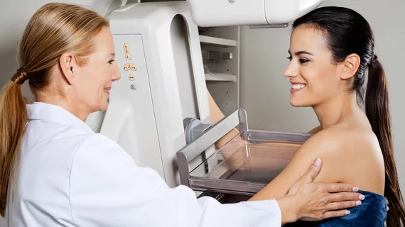
Follow-up imaging for women with non-metastatic breast cancer differs widely across the U.S., research out of the University of California, San Francisco, has found. The key factor in discerning patients' follow-up treatment seems to be where they live.
Benjamin Franc, MD, MS, an investigator for the study and radiology professor at UCSF, said in a release from UCSF he and his team examined data from 36,045 women between 18 and 64 years old who had surgery for cancer in one breast between 2010 and 2012. All patients presented with non-metastatic disease.
During an 18-month study period that allowed patients time to complete any radiation therapy they needed after surgery, Franc said the researchers found that 70.8 percent of women had received at least one dedicated breast image—an MRI or a mammogram—during that time, while 31.7 percent received at least one high-cost imaging procedure. Some 12.5 percent had at least one PET scan, which isn’t recommended in non-metastatic cases.
The American Society of Clinical Oncology and the National Comprehensive Cancer Network recommend women with non-metastatic disease receive annual physical exams and mammograms, but not full-body imaging with CT, MRI or PET.
“These patients already have cancer, so you don’t want to induce another cancer with radiation from unnecessary imaging,” Franc said in the release.
According to the study, patients were more likely to undergo recommended breast screening within 18 months of surgery if they were younger or had received radiation therapy. The next most common predictor, the patient’s metropolitan statistical area, surprised Franc.
“Age and therapy make sense as predictors of breast imaging, but it doesn’t make sense that where you live makes a difference in whether you were likely to get a follow-up mammogram or high-cost imaging,” he said. “What’s actionable here is that we have these guidelines, but doctors aren’t following them.”
Depending on where the women lived, Franc said, between 18 and 46 percent of patients received high-cost tomographic imaging within a year and a half of surgery. And that’s not an affordable price tag for a sizeable fraction of breast cancer patients, some of whom have to pay up to $8,000 in deductible fees every year. A full-body scan alone costs between $2,000 and $8,000.
“With skyrocketing medical costs, patients are having to take greater and greater responsibility for out-of-pocket expenses,” Franc said. He said his team couldn’t find any other patterns in their data to explain the wide variation in post-surgery breast imaging.
Franc et al.’s research was published Friday in the Journal of the National Comprehensive Cancer Network.

