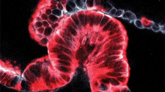
Mystery solved: New imaging technique a game-changer for pancreatic cancer treatment
A new 3D imaging technique has provided researchers with insight into how pancreatic cancers start and grow, according to findings published in Nature. It’s a discovery that could have a significant impact on patient care going forward.
For decades, scientists have struggled to make sense of how pancreatic tumors start. 2D slices just weren’t providing enough information. But thanks to this breakthrough, which involves the imaging of tissue biopsies, it is now evident that two types of cancer are forming in these ductal cells: “endophytic” tumors, which grow into the ducts, and “exophytic” tumors, which grow outward.
“To investigate the origins of pancreatic cancer, we spent six years developing a new method to analyze cancer biopsies in three dimensions,” Hendrik Messal, a researcher at the Francis Crick Institute in London and co-lead author of the Nature study, said in a prepared statement. “This technique revealed that cancers develop in the duct walls and either grow inwards or outwards depending on the size of the duct. This explains the mysterious shape differences that we’ve been seeing in 2D slices for decades.”
The researchers also used this new 3D imaging technique on other organs, noting that cancers in the lungs and liver act in the same way.
“This technological breakthrough has the potential to unlock many unanswered questions of great importance in how we understand and treat pancreatic cancer,” Andrew Biankin, a professor who serves as Cancer Research UK’s pancreatic cancer expert, said in the same prepared statement. “It’s crucial we better grasp how these cancers behave from the earliest stages, to help develop treatments for a disease where survival rates have remained stubbornly low.”
Two different research groups at the Francis Crick Institute worked together on this project. Support also came from the European Research Council, Cancer Research UK, the Medical Research Council and Wellcome.

