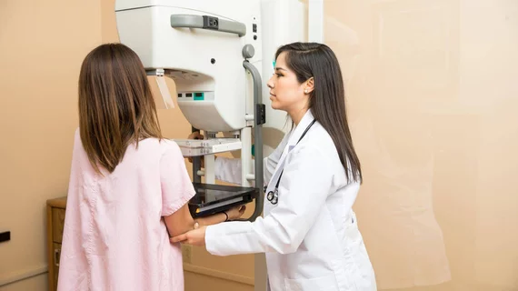
Researchers study the effectiveness of self-compression during mammography
Self-compression during mammography does not lead to a rise in patient discomfort or a drop in image quality, according to new findings published in JAMA Internal Medicine.
“Despite its utility, many women dread undergoing mammography because this examination can be uncomfortable or painful,” wrote Philippe Henrot, MD, department of radiology at the Institut de Cancérologie de Lorraine Alexis Vautrin in France, and colleagues. “This apprehension has been observed in the field of screening; pain or discomfort experienced by women has been reported as a common factor in nonattendance or non-reattendance of screening.”
The authors noted that numerous techniques have been studied to address this issue and help more women receive care. One of those techniques is self-compression, when the patient is given control of the compression of her own breasts during the mammogram. To explore the effectiveness of that technique, the team studied data from mammograms conducted from May 7, 2013, to Oct. 26, 2015, at six cancer care centers in France. All patients were between the ages of 50 and 75, physically able to perform self-compression and had no recent history of breast surgery or treatment.
While 275 patients were included in the self-compression group, 273 were included in the traditional compression group. The authors found that the difference in mean breast thickness between the two groups’ mammography results was “lower than the noninferiority margin” and compression force was higher in the self-compression group. In addition, the self-compression group reported less pain, and there was no difference in image quality.
“Self-compression does not appear to be inferior to standard compression in achieving minimal breast thickness,” the authors wrote. “With this technique, a higher compression force is achieved without increasing pain or compromising image quality. Self-compression mammography may be an effective screening approach for women who want to take an active role in their breast examination.”
The research did have some limitations, including the fact that radiation dose was not evaluated. In addition, the team noted, the amount of time it took for self-compression to occur was not measured.

