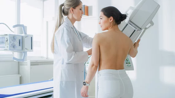
Researchers receive $3.6M grant to develop less painful cancer detection tool
University of Arizona Health Sciences scientists are developing a new 3D breast-specific cancer detection method that would do away with the often-painful compression methods used during mammograms.
Their new CT tool could also provide an accurate estimate of breast density and reduce false positives.The National Cancer Institute is helping to fuel such innovation with a $3.6 million five-year grant, the school announced.
University of Arizona President Robert Robbins, MD, said this research dovetails perfectly with the school’s strategic plan.
“This research to develop new technology for breast cancer imaging, followed by a clinical trial, is a great example of how we should use the university’s strengths as an innovation powerhouse to create positive impact for patients, their families and communities around the world,” he said in a prepared statement.
Andrew Karellas and Srinivasan Vedantham—two professors in the department of medical imaging who have a passion for detecting breast cancer as early as possible—will lead the effort. Their team hopes to eventually develop and clinically evaluate the next generation of breast CT. Researchers believe the 3D nature of their tomographic images will improve detection and help physicians determine whether findings are malignant or noncancerous more quickly.

