
Deep learning shines at predicting incidental lung nodules' severity
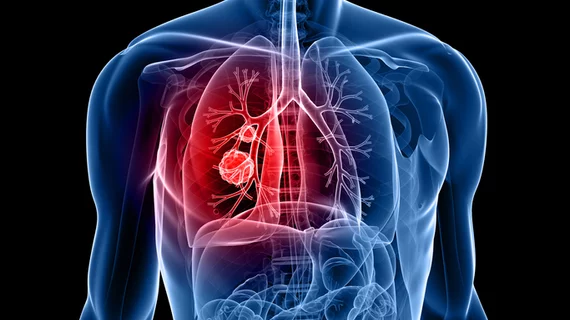
Researchers have developed deep learning models that outperform current ones at predicting whether incidental lung lesions will become malignant.
A new paper in European Radiology details the development and testing of two DL models that combine imaging and clinical data to calculate a lesion's probability of malignancy. When externally validated on a diverse test set of scans from numerous institutions, the models yielded greater sensitivity and specificity than the Brock model, a widely used malignancy prediction tool that combines imaging and patient features through predefined variables. Experts believe the models’ performance on a more diverse dataset gives them an edge over other prediction alternatives in real world settings.
One of the models was trained using screening data, while an updated version incorporated both screening and clinical data. Researchers sought to determine whether the models could maintain their performance across different centers with varying equipment and whether adding clinical data would improve their predictive ability. The retrospective multicenter study included 269 incidental pulmonary nodules from 231 patients. The nodules were stratified by size, with 5–10 mm, 10–15 mm and 15–30 mm categories included.
Of the nodules, 89 were malignant. The screening-trained DL model achieved an AUC of 0.74, while the model trained on both screening and clinical data yielded an AUC of 0.72; both outperformed the Brock model, which had an AUC of 0.63. This difference also was evident when the models were evaluated at a fixed sensitivity; using a 10% threshold for the Brock model, both DL models achieved 60% specificity compared with 44% for the Brock model. These differences were maintained across three participating institutions, with the Brock model consistently being outperformed by the other two.
“This study provides evidence of consistent model performance across centers with varying CT and patient populations within one country, providing further evidence of the potential of DL models to classify pulmonary nodules in varying circumstances,” Renate Dinnessen, of the department of medical imaging at Radboud University Medical Center in the Netherlands, and colleagues concluded. “Future work should look at how to integrate such DL malignancy probability scores into management protocols, the interaction between these models and clinicians, and the acceptance of such models, as within a clinical setting, these DL models could assist the clinician.”
Read more here.

In addition to her background in journalism, Hannah also has patient-facing experience in clinical settings, having spent more than 12 years working as a registered rad tech. She began covering the medical imaging industry for Innovate Healthcare in 2021.