Cardiac Imaging

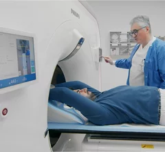
While cardiac ultrasound is the widely used imaging modality for heart assessments, computed tomography (CT), magnetic resonance imaging (MRI) and nuclear imaging are also used and are often complimentary, each offering specific details about the heart other modalities cannot. For this reason the clinical question being asked often determines the imaging test that will be used.
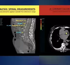
The new cardiac PET radiotracer flurpiridaz F-18 is posed to be a major game changer and will likely lead to increased adoption of cardiac PET.

FDA approves GE HealthCare's flurpiridaz F-18 PET radiotracer for CAD

The newly approved radiotracer is seen as a major step forward for nuclear imaging technology. Specialists have been looking forward to its arrival on the market for quite some time.

Time running out to comment on higher cardiac CT payments

Imaging experts have said the move could reshape the financial landscape for hospitals offering Cardiac CT services.

Example of AI automated detection and highlighting of critical lung findings on a chest X-ray for a possible lung cancer nodule and fibrosis. Example shown by AI vendor Lunit.

PHOTO GALLERY: Examples of FDA-cleared AI in radiology

This is a photo gallery of artificial intelligence products cleared for clinical use in medical imaging by the U.S. Food and Drug Administration. Radiology by far is the leader of all clinical AI FDA approvals.

Ron Blankstein, MD, FACC, FASNC, MSCCT, FASPC, associate director, cardiovascular imaging program, director, cardiac computed tomography, Brigham and Women’s Hospital, and a professor of medicine and radiology, Harvard Medical School, explains why Medicare is proposing increased coronary computed tomography angiography (CCTA) payments to hospitals.

Higher payments for CCTA could bring considerable change to cardiology

Ron Blankstein, MD, explains how and why Medicare is proposing an increase to hospital payments for coronary CT angiography.

CMS may double Medicare payments for cardiac CT

The agency is gathering feedback on a proposal to double the amount hospitals are paid for coronary computed tomography angiography, with the comment period ending Sept. 9.

The American Society of Nuclear Cardiology (ASNC) is asking Congress to repeal the appropriate use software provision mandate, which physicians say is an obstacle to efficient care.

ASNC asks Congress to officially repeal the AUC mandate for advanced medical imaging

Medicare rescinded the provision in the 2024 Medicare Physician Fee Schedule, but the law remains on the books. 

How the proposed Medicare Physician Fee Schedule could impact nuclear cardiology

The American Society of Nuclear Cardiology explored some key points included in the 2025 Medicare Physician Fee Schedule proposed rule.

Researchers with University College London and the European Synchrotron Radiation Facility (ESRF) have used a new X-ray technique, hierarchical phase-contrast tomography (HiP-CT), to capture images of the human heart in unprecedented detail. The group shared its images, as well as a full analysis, in Radiology.[1]

New imaging technique captures human heart with 'unprecedented detail'

The new images were captured at the European Synchrotron Radiation Facility using hierarchical phase-contrast tomography. One specialist called them "Google Earth for the human heart." 

Around the web

The new F-18 flurpiridaz radiotracer is expected to help drive cardiac PET growth, but it requires waiting between rest and stress scans. Software from MultiFunctional Imaging can help care teams combat that problem.

News of an incident is a stark reminder that healthcare workers and patients aren’t the only ones who need to be aware around MRI suites.

The ACR hopes these changes, including the addition of diagnostic performance feedback, will help reduce the number of patients with incidental nodules lost to follow-up each year.