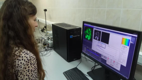
Modern FLIM system allows for high-res imaging on a macro scale
Scientists in Germany and Russia have achieved macroscale imaging with fluorescence lifetime imaging microscopy (FLIM), opening the field to opportunities for imaging whole tumors and areas the size of several square centimeters, the Optical Society announced this week.
Research led by Vladislav Shcheslavskiy, PhD, of Becker & Hickl in Germany, found FLIM could be scaled up to create the first-ever confocal microscopy-based macro-FLIM system using ultrasensitive detectors and picosecond lasers. Shcheslavskiy’s team tested the technique in fluorescent microbeads, live cultured cancer cells and a full tumor in a live mouse—none of which are possible with current FLIM capabilities.
“Our macro-FLIM system can not only obtain a sample’s structural information, but also allows observation of certain biochemical processes taking place within the sample,” Shcheslavskiy said in a release from the OSA. “Although our goal is to develop this for clinical use, it could also be very useful in fundamental studies for probing biological processes as disease develops or investigating biological responses to different types of therapy.”
The system uses laser scanning confocal microscopy to gather information about a sample’s properties and its micro-environment. High-resolution imaging was attained by incorporating short-pulse lasers, which, combined with sensitive detectors, are able to sense fluorescence.
“Careful optical design along with the picosecond lasers, sensitive and fast detectors and fast single photon counting electronics allows us to record fluorescence decay with high precision at macroscale,” Shcheslavskiy said, noting the macro-FLIM system can analyze molecular samples up to 4 square centimeters in area. Typically, FLIM can only measure spaces a few millimeters wide.
To analyze the mouse tumor, Shcheslavskiy said the researchers had to simultaneously measure the fluorescence lifetime of a genetically encoded red fluorescent protein, which identified the location of the tumor, and nicotinamide adenine dinucleotide.
Shcheslavskiy said the team’s ultimate goal is to bring macro-FLIM to clinical practice and use the method to identify the edges of tumors during surgery, but it could also be applied to other areas. For example, he said, it could be used as a non-destructive method for identifying materials in paintings that need restoring.
“The sensitivity of our system was high enough to observe fluorescence of intrinsic tissue components such as NADH without any labeling,” he said. “In addition to being used to study metabolism in a tumor, macro-FLIM could be used to follow cell death or oxygen status of tumors on a macroscale with cellular resolution.”

