Pulmonary embolism

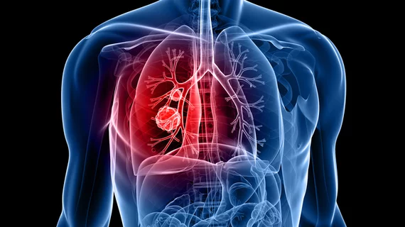
Pulmonary embolism (PE) is the third leading cardiovascular cause of death after heart attacks and stroke. PE is caused by blood clots in the pulmonary arteries. These are often caused by clots from the venous system, including thrombus from trauma, surgery or deep vein thrombosis (DVT). Treatment has traditionally been systemic use of thrombolytic drugs to dissolve the clot. But in cases there is a massive, life-threatening PE, or chronic clot burden that have remained in a vessel for an extended period of time, mechanical thrombectomy and ultrasound-assisted catheter-directed thrombolysis (USCDT) is being used as more targeted and aggressive treatments.

Thumbnail

RSNA offering cash, massive dataset for AI solutions to tackle pulmonary embolism

The Radiological Society of North America launched its fourth annual artificial intelligence challenge, hoping to help docs detect and characterize the condition. 

Thumbnail

Catheter-directed therapy treatments for pulmonary embolism on the rise

Catheter-directed therapy (CDT) is being used to treat pulmonary embolism (PE) more and more by healthcare providers, according to new research published in the Journal of Vascular and Interventional Radiology.

Thumbnail

AI solution for pulmonary embolism workflow receives CE certification

Aidoc, a Tel-Aviv, Israel-based medical imaging company, announced that its AI-based solution for improving pulmonary embolism (PE) workflow has received CE certification.

Thumbnail

IVC filters improve in-hospital mortality rates for patients with congestive heart failure, pulmonary embolism

Inferior vena cava (IVC) filter placement is associated with improved in-hospital mortality rates for patients admitted with congestive heart failure (CHF) and pulmonary embolism (PE), according to a new study published by the American Journal of Roentgenology.

Example of spectral, or dual-energy CT, confirming a pulmonary embolism (PE). Image courtesy of Philips Healthcare

MRA vs CTA for evaluating pulmonary embolism: Does the chosen modality impact downstream imaging utilization?

MR angiography (MRA) is a relatively new alternative to CT angiography (CTA) for the evaluation of suspected pulmonary embolism (PE) and it even has one major advantage over CTA: it does not expose patients to ionizing radiation.

Thumbnail

Chest CT for suspected pulmonary embolism in the ED: Utilization is up, diagnostic yield is down

CT utilization in the emergency department (ED) has increased significantly in the last few decades, a fact often cited as one of the major reasons healthcare costs and radiation exposure are on the rise. So what can be done to reverse that trend?

pulmonary embolism on CT pulmonary angiography

Feel free to reduce CT dose when diagnosing pulmonary embolism

Clinicians can reduce the “tube voltage” (kVp) in CT pulmonary angiography without reducing image quality, according to a study published in the Journal of the American College of Radiology. Researchers conducted a retrospective study to measure CT pulmonary angiography image quality as tube voltage was lowered, taking into account patient size as well as the benefits of simple image post-processing.

Intrinsic Imaging awarded interventional medical device trial for prevention of pulmonary emboli

Intrinsic Imaging, an FDA audited, ISO 9001:2008 and ISO 13485:2003 certified, GAMP 5 compliant medical imaging core lab specializing in providing services for clinical trials, announced today that it has been awarded a clinical trial to study an interventional medical device designed for the prevention of pulmonary emboli.

Around the web

The patient, who was being cared for in the ICU, was not accompanied or monitored by nursing staff during his exam, despite being sedated.

The nuclear imaging isotope shortage of molybdenum-99 may be over now that the sidelined reactor is restarting. ASNC's president says PET and new SPECT technologies helped cardiac imaging labs better weather the storm.

CMS has more than doubled the CCTA payment rate from $175 to $357.13. The move, expected to have a significant impact on the utilization of cardiac CT, received immediate praise from imaging specialists.