
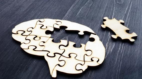
MRI findings suggest parts of the brain reorganize to become 'younger' after a stroke
New MRI findings detail how the brain compensates for stroke-related damage by reorganizing unaffected areas.
Published in The Lancet Digital Health, the study suggests that when certain areas of the brain are damaged, other regions opposite the lesion appear to decelerate ageing. The finding could signal that the brain has the potential to heal itself in the long-term after a stroke.
“We found that larger strokes accelerate aging in the damaged hemisphere but paradoxically make the opposite side of the brain appear younger,” noted co-senior author of the study Hosung Kim, PhD, associate professor of research neurology at the Keck School of Medicine of USC. “This pattern suggests the brain may be reorganizing itself, essentially rejuvenating undamaged networks to compensate for lost function.”
For the study, experts analyzed the MRI scans of 500 stroke survivors across 34 research sites in eight countries. Deep learning techniques that were trained on tens of thousands of brain scans were harnessed to estimate the “brain age” of 18 different regions of the brain to get a better idea of how those affected by a stroke compared to healthy regions. Motor performance scores were used to gauge participants’ level of impairment.
Through this the team observed a pattern: participants with worse motor performance scores showed younger-than-expected brain ages in areas opposite of where the lesion was located. This finding was notably prominent in the frontoparietal network, which is known to be associated with motor function, attention and coordination. This suggests that the opposite hemisphere of the affected side of the brain is capable of compensating for stroke damage, the authors signaled.
“We saw this in the contralesional frontoparietal network, which showed a more ‘youthful’ pattern and is known to support motor planning, attention, and coordination. Rather than indicating full recovery of movement, this pattern may reflect the brain’s attempt to adjust when the damaged motor system can no longer function normally,” Kim said. “This gives us a new way to see neuroplasticity that traditional imaging could not capture.”
The group has plans to continue their work in longitudinal studies to track how these patterns change in the long-term. They are hopeful that their work could lead to more personalized management for stroke patients.
Read more here.

In addition to her background in journalism, Hannah also has patient-facing experience in clinical settings, having spent more than 12 years working as a registered rad tech. She began covering the medical imaging industry for Innovate Healthcare in 2021.