Magnetic Resonance Imaging

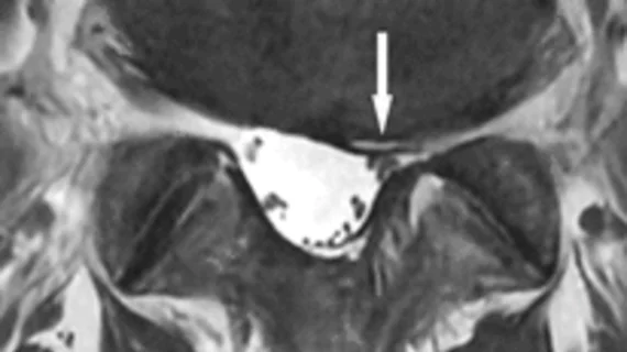
Magnetic resonance imaging (MRI) is the gold standard imaging modality for soft tissues. It produces detail cross-sectional images of soft tissue and bone anatomy, including muscles, tendons, ligaments, brain and organs, without the use of ionizing radiation. In addition to orthopedic imaging, MRI is also used for heart, brain and breast. MRI uses gadolinium contrast in many exams to highlight tissues and blood vessels, which enhances images and offers better diagnostic quality. It can also be used in conjunction with PET scans. How does MRI work? MR creates images by using powerful magnets to polarize hydrogen atoms in water (the body is made of of more than 80% water) so they face in one direction. A radiofrequency pulse is then used to ping these atoms, causing them to wobble, or resonate. The MRI coils detect this and computers can assemble images from the signals. Basic MRI scans will focus on the resonance of fat and water in two different sequences, which highlight and contrast different features in the anatomy.

Strategic MRI Acquisition Grows Volume: Imaging Healthcare Specialists

Sponsored by Hitachi Healthcare Americas

With 26 radiologists on staff and 11 locations in the San Diego, California, area, Imaging Healthcare Specialists (IHS) is the largest provider of outpatient imaging services in San Diego county; its reach extends into neighboring Riverside County as well. Like any busy radiology practice, IHS has a strong focus on MRI services, with 13 MRI systems

Functional MRI May Be Early Autism Indicator

Functional magnetic resonance imaging (fMRI) may serve as an early and objective indicator of autism, according to a study slated for publication in the August issue of Radiology. Already posted online, the study was conducted by researchers at Columbia University in New York City, who used the technique to document language impairment in autistic

MR Knee Coil Receives FDA Clearance

The U.S. Food and Drug Administration (FDA) has cleared for use Toshiba America Medical Systems’ extra-large knee array coil for the Vantage TitanTM MR system. Toshiba claims to be the first diagnostic imaging vendor to offer an extra-large knee coil with a 22-cm interior diameter for MR imaging.

Algorithm Improves MRI Interpretation

An improved algorithm can dramatically enhance the capture and interpretation of full-body MRIs, particularly in the abdominal region, according to a new study slated for presentation Thursday at the annual meeting of the American Roentgen Ray Society (ARRS) in Chicago.

Self-Referring Physicians Order More MRI Scans

Physicians who own or lease MRI equipment are more likely than those who do not have such equipment in-house to perform scans on patients with low back pain, according to a study performed at Stanford University. Results of the study, published online yesterday in Health Services Research, also indicated that patients whose orthopedists perform MRI

RIS-driven Workflow: Enhanced Clinical History Through the EMR

Sponsored by FUJIFILM Healthcare Americas

Access to clinical history is of utmost importance to the radiologists of The Children’s Hospital in Denver, Colorado. Because the hospital’s main campus and five satellite sites generate around 130,000 pediatric images a year, it’s critical for radiologists to be able to access a patient’s indications and background quickly, while maintaining the

MRI Offers No Added Value In RA Risk Stratification

MRI is no more cost-effective than standard risk stratification techniques in determining whether newly diagnosed rheumatoid arthritis (RA) patients will benefit from aggressive treatment is no more cost-effective than standard risk stratification techniques, according to researchers at the Yale School of Medicine in New Haven, Conn.

MR Systems Garner FDA Clearance

GE Healthcare yesterday announced FDA clearance of its Optima™ MR360 1.5T and Brivo™ MR355 1.5T magnetic resonance systems. Approximately 180 such systems have been ordered by hospitals and imaging centers worldwide.

Around the web

The nuclear imaging isotope shortage of molybdenum-99 may be over now that the sidelined reactor is restarting. ASNC's president says PET and new SPECT technologies helped cardiac imaging labs better weather the storm.

CMS has more than doubled the CCTA payment rate from $175 to $357.13. The move, expected to have a significant impact on the utilization of cardiac CT, received immediate praise from imaging specialists.

The all-in-one Omni Legend PET/CT scanner is now being manufactured in a new production facility in Waukesha, Wisconsin.