Magnetic Resonance Imaging

Magnetic resonance imaging (MRI) is the gold standard imaging modality for soft tissues. It produces detail cross-sectional images of soft tissue and bone anatomy, including muscles, tendons, ligaments, brain and organs, without the use of ionizing radiation. In addition to orthopedic imaging, MRI is also used for heart, brain and breast. MRI uses gadolinium contrast in many exams to highlight tissues and blood vessels, which enhances images and offers better diagnostic quality. It can also be used in conjunction with PET scans. How does MRI work? MR creates images by using powerful magnets to polarize hydrogen atoms in water (the body is made of of more than 80% water) so they face in one direction. A radiofrequency pulse is then used to ping these atoms, causing them to wobble, or resonate. The MRI coils detect this and computers can assemble images from the signals. Basic MRI scans will focus on the resonance of fat and water in two different sequences, which highlight and contrast different features in the anatomy.

Thumbnail

AI looking handy with 3D abdominal ultrasound

Mayo Clinic researchers have demonstrated a deep learning model that can automatically segment kidneys and measure total kidney volumes using only 3D ultrasound images.

Thumbnail

Abbreviated breast MRI deemed an attractive screening option—sometimes

AB-MRI is a cost-effective means of screening women with dense breast tissue for breast cancer—as long as the per-exam costs don’t top 82% of what would have been spent to perform full-protocol breast MRI.

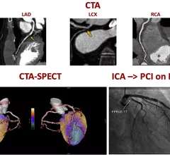
A figure from the 2022 CAD non-invasive imaging guidelines showing a comparison of computed tomography angiography (CTA) and a SPECT-CT vs. an invasive angiogram from the cath lab showing the same blockage in a coronary artery.

New multi-society recommendations highlight role of non-invasive imaging in evaluating coronary artery disease

A new, multi-society document, "Non-Invasive Imaging in Coronary Syndromes," focuses on how multiple imaging techniques can evaluate different aspects of coronary artery disease (CAD), all without the need for invasive angiograms.

Thumbnail

Use of unnecessary neuroimaging for patients with dizziness prevalent in outpatient settings

Quality improvement efforts have sought to address such low-value diagnostic testing in the ED, with little attention paid to ambulatory settings, experts wrote in JAMA

Private equity-backed Rayus Radiology details latest expansion effort

The Minneapolis-based imaging group is broadening its partnership with Froedtert & the Medical College of Wisconsin, leaders announced recently. 

quality imaging appropriateness clinical decision support CAS AUC

Application for reporting adverse contrast reactions significantly increases radiologists’ thoroughness

Mass General has piloted the use of a new tool called CISaR (Contrast Incident Support and Reporting) to vastly improve documentation.

button battery

MRI after button battery removal: Consensus on next steps lacking among referrers

There are no published guidelines on how imaging should inform clinical management, Seattle Children's Hospital experts noted. 

quality

Study charts uptick in unnecessary CT, MR imaging surveillance of noninvasive bladder cancer

The impact of these patterns is substantial and may have negative consequences for patients and the healthcare system, experts wrote in JAMA Network Open

Around the web

The nuclear imaging isotope shortage of molybdenum-99 may be over now that the sidelined reactor is restarting. ASNC's president says PET and new SPECT technologies helped cardiac imaging labs better weather the storm.

CMS has more than doubled the CCTA payment rate from $175 to $357.13. The move, expected to have a significant impact on the utilization of cardiac CT, received immediate praise from imaging specialists.

The all-in-one Omni Legend PET/CT scanner is now being manufactured in a new production facility in Waukesha, Wisconsin.

Trimed Popup
Trimed Popup