
Fluorescent contrast agent noninvasively identifies common skin cancer
A fluorescent contrast agent could improve the diagnosis of certain skin cancers, according to new research detailed in the Journal of Nuclear Medicine.
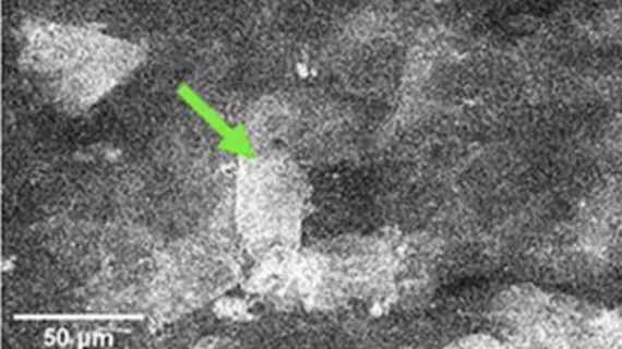
PARPi-F is a poly(adenosine diphosphate ribose) polymerase 1 (PARP1) inhibitor–targeted contrast agent. When used topically, it penetrates the skin and generates a fluorescent signal under the guidance of a fluorescent confocal microscope (FCM). Researchers believe that its use can improve the diagnosis and treatment of basal cell carcinoma—the most common type of skin cancer.
BCC is highly treatable when it is identified in its early stages before it has penetrated into the deeper layers of skin. However, BCC diagnoses can sometimes be a lengthy process, which prolongs the time-to-treatment, and the process can leave patients with unwanted scars. As such, there is an interest in improving the diagnostic process and making it more patient-friendly and timely.
“Emerging nonsurgical therapies for early basal cell carcinomas could be delivered at the bedside, but they require noninvasive diagnostic tools with high accuracy,” explained Manu Jain, MD, research pathologist and optical imaging specialist, dermatology, at Memorial Sloan Kettering Cancer Center in New York. “Our study investigated the feasibility of using fluorescent confocal microscopy with PARPi-FL for enhancing basal cell carcinoma detection in the clinical setting.”
For the research, the team analyzed the effect of PARPi-F on ex vivo human tissue samples acquired during plastic and Mohs surgeries; they also conducted an in vivo analysis using an FCM on tumor-bearing mice. A topical dose of 10 µM was applied to the samples via gauze for 2 to 5 minutes to ensure penetration of the substance.
PARPi-FL achieved sufficient dermal penetration, yielding a strong fluorescent signal. These signals were much stronger in basal cell carcinomas compared to benign lesions. Additional toxicity assessments indicated the agent was safe for topical use as well.
“Incorporating this targeted dye into in vivo imaging could significantly improve diagnostic accuracy, reduce unnecessary biopsies of benign lesions, and enable timely, noninvasive treatment for basal cell carcinoma,” Jain said.
The team added that PARP1 is also overexpressed in melanomas, suggesting the contrast agent could have utility beyond basal cell carcinoma in the future.
Learn more about the findings here.

In addition to her background in journalism, Hannah also has patient-facing experience in clinical settings, having spent more than 12 years working as a registered rad tech. She began covering the medical imaging industry for Innovate Healthcare in 2021.