
Whole-body PET system cuts scan time by over 80%
Experts believe new developments in whole-body PET scanners have the potential to significantly reduce scan times and improve imaging workflows.
In fact, a new paper published in the Journal of Nuclear Medicine suggests that these scanners can slash scan times by as much as 83%. And the saved time does not come at the expense of image quality—it may actually increase lesion detection instead.
The study compares the use of a long-axis field-of-view (LAFOV) PET/CT scanner to conventional lutetium oxyorthosilicate-based (LSO/LYSO) short-axial field-of-view (SAFOV) scanners, analyzing both its timesaving and lesion detection capabilities. Short-axial scanners are the standard of care, but long-axis alternatives have gained traction in recent years due to their ability to provide whole-body imaging without requiring patients to change positions throughout an exam.
“Long-axis field-of-view PET/CT offers increased sensitivity that may enable shorter acquisition time while maintaining or improving diagnostic image quality,” lead study author Alicia Corlett and co-authors noted.
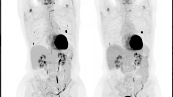
The team recently recruited a group of oncology patients to undergo a dual scanning protocol following a single administration of either 18F-FDG, 1F-DCFPyL, 68Ga-DOTATATE or 68Ga-PSMA. Patients first completed an exam using a short-axial scanner (GE HealthCare Discovery 710 or Siemens Healthineers Vision 600) before completing an additional scan immediately after on a long-axis scanner (Omni 128 cm PET/CT, GE HealthCare) using a single 10-min list-mode acquisition, with rebinning to match short-axial, per-bed acquisition times for direct comparison. Three nuclear medicine specialists assessed the exams for image quality.
Exams on the long-axial scanner took just 2.5 minutes, while short-axial one took around 15. Long-asix images also scored consistently higher across multiple measures of quality, including noise, sharpness and lesion conspicuity. The long-axial exams increased lesion detection as well, with 68% of patients having additional smaller lesions identified compared to what was visible on their short-axis scan. These findings remained consistent across the different radiotracers.
Some of the differences between the exams could potentially be partially attributable to the different detector materials; long-axial detectors are made from germanium oxide with silicon photomultipliers, the authors explained.
“LAFOV PET/CT provides superior image quality compared to SAFOV PET/CT despite marked reductions in acquisition time, with comparable noise characteristics across multiple radiopharmaceuticals,” the team wrote.
The study abstract is available here.

In addition to her background in journalism, Hannah also has patient-facing experience in clinical settings, having spent more than 12 years working as a registered rad tech. She began covering the medical imaging industry for Innovate Healthcare in 2021.