
Providers must tread carefully when using shortened PSMA PET/CT protocols
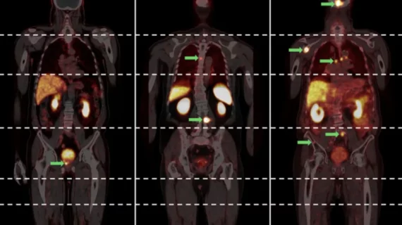
A more limited imaging protocol may be sufficient to detect relevant prostate cancer recurrence in many patients undergoing prostate-specific membrane antigen (PSMA) PET/CT, potentially reducing scan time and resource use.
However, this runs the risk of missing lesions in some patients, warranting judicious use, according to a new paper published in BMC Medical Imaging. This is something that providers must weigh carefully when deciding which patients may be eligible for shortened protocols.
“In analogy to other PET/CT applications, PSMA-PET/CT is commonly performed as a whole-body examination. This contrasts with conventional radiological imaging modalities such as CT or MRI, which are typically tailored to specific anatomical regions based on the clinical question,” Martin Freesmeyer, with the clinic of nuclear medicine at University Hospital Jena, Germany, and colleagues noted. “Considering metastatic mechanisms and the characteristic pathways of [prostate cancer] tumor spread, which usually involve the prostate bed, locoregional lymph nodes and the axial skeleton, while distal metastases—especially in the lower extremities—are rare, the necessity of extended whole-body imaging warrants reconsideration.”
To support their conclusions, researchers conducted a retrospective, single-center study that included 200 patients with biochemically recurrent prostate cancer who underwent gallium-68 PSMA PET/CT imaging from the vertex to the toes. The team compared the findings with hypothetical limited scan protocols, including vertex-to-mid thigh, skull base-to-mid thigh, chest-abdomen-pelvis, abdomen-pelvis and pelvis-only coverage.
Overall, 128 patients (64%) had at least one structural correlate of recurrent prostate cancer on whole-body imaging. This rate carried over to the two limited protocols commonly used in clinical practice and with chest-abdomen-pelvis imaging as well.
However, narrowing the scan area further resulted in missed disease. A chest-abdomen-pelvis protocol would have failed to identify all tumors in 16 patients (8%) and all affected organ systems in six patients (3%). An abdomen-pelvis protocol would have missed signs of recurrence in 41 patients (21%), while pelvis-only imaging would have missed them in 61 patients (31%).
Prostate-specific antigen levels appeared to be beneficial in identifying patients who could undergo shortened imaging protocols. Among patients with PSA levels of 0.75 ng/mL or lower, researchers found no additional diagnostic benefit from whole-body coverage. Based on this threshold, approximately 45% of the study population could have undergone chest-abdomen-pelvis imaging without losing diagnostic information.
These findings suggest that tailoring PSMA PET/CT coverage based on PSA levels may offer a practical way to balance imaging efficiency without the risk of missing metastatic disease.
“Due to restricted availability, an optimization of scanning protocols is necessary to make this method accessible for every patient fulfilling its evidence-based indication. Based on the results of this study, we do not recommend true whole-body scans for patients without specific reasons,” the group noted. “A standard scan from vertex to mid-thigh seems sufficient without missing relevant metastases. In cases with low PSA levels (i.e., ≤ 0.75 ng/mL), an even more focused protocol of only chest/abdomen/pelvis can be considered.”
Read more here.

In addition to her background in journalism, Hannah also has patient-facing experience in clinical settings, having spent more than 12 years working as a registered rad tech. She began covering the medical imaging industry for Innovate Healthcare in 2021.