
Targeted immunoPET imaging detects multiple myeloma missed by standard PET/CT
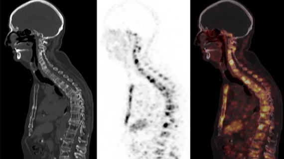
A targeted immunoPET scan can identify active multiple myeloma in patients whose disease is often missed by conventional PET/CT imaging.
That’s according to a new a multicenter phase 2 clinical trial led by researchers at the University of Miami's Sylvester Comprehensive Cancer Center. The study, published in RSNA's Radiology, evaluated an antibody-based imaging approach designed to bind to a protein expressed on the surface of multiple myeloma cells known as CD38. Researchers labeled the antibodies with a radioactive tracer to make cancerous cells visible on PET imaging, offering an alternative to conventional scans that rely on glucose uptake to identify tumors.
Standard glucose-based PET/CT can miss multiple myeloma because some cancer cells consume glucose at levels similar to surrounding normal tissue. It is estimated that these conventional scans fail to identify residual disease in as many as 40% of cases.
“If we are going to cure multiple myeloma, we need two things. One, we need really effective medicines. Second, we need to be able to make sure there is no disease left behind after these treatments,” C. Ola Landgren, MD, PhD, director of the Sylvester Myeloma Institute, and senior author of the study, said in a news announcement on the findings. “It's very well known among radiologists and myeloma experts that some patients who have myeloma in their bones may not light up on PET scans. The methods are simply not designed to reliably capture multiple myeloma.”
The trial included 60 patients with multiple myeloma who underwent both conventional PET/CT and the targeted immunoPET exam. ImmunoPET identified disease not detected by standard imaging in 17 patients (28%); in 13 patients (22%), conventional PET/CT identified disease that immunoPET missed. The two approaches detected a similar amount of disease in the remaining participants. However, follow-up biopsies confirmed active myeloma in areas identified by immunoPET but not conventional imaging.
In one example, a patient with persistent back pain had normal blood markers and unremarkable standard imaging, while immunoPET revealed spinal lesions. Another patient whose initial scans and blood tests showed no evidence of disease had nearly 100 sites of suspected myeloma identified with the targeted approach, with biopsy confirming the findings.
The results suggest immunoPET could provide complementary information to conventional diagnostic pathways when assessing disease burden and evaluating treatment response. However, the findings also demonstrate that neither imaging approach is foolproof, as both missed some cases. This supports the value of combining diagnostic methods rather than replacing existing tests, the authors noted. Further studies will be needed to establish the technology's clinical utility and determine whether it improves treatment decisions or patient outcomes.
Read more here.

In addition to her background in journalism, Hannah also has patient-facing experience in clinical settings, having spent more than 12 years working as a registered rad tech. She began covering the medical imaging industry for Innovate Healthcare in 2021.