
Imaging technique records seizures in real time
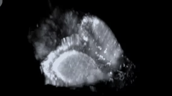
University of Georgia researchers have developed a high-resolution light-sheet imaging system capable of capturing seizures moving through the brain in three dimensions and in real time.
The imaging system combines light-sheet microscopy with adaptive optics. Light-sheet microscopy uses a thin sheet of light to illuminate individual slices of a sample, allowing researchers to acquire high-speed images with reduced background noise. This approach is helpful for observing biological processes in living organisms. Adaptive optics further improve image quality by correcting distortions caused as light travels through biological tissue.
“When you look at the stars in the night sky, they sort of twinkle because the atmosphere is making the image wobble around,” corresponding author Peter Kner, PhD, a professor at the UGA College of Engineering, said in a news release Aug. 12. “Adaptive optics technology corrects that.”
Applying the same principle to brain imaging allows the microscope to produce sharper images despite the optical distortions created by tissue. Achieving this in 3D instead of 2D could offer new insight into how seizure activity begins and spreads throughout its duration.
“The brain is obviously three dimensional, so when you have 2D imaging, not everything is going to be visible on that single 2D plane,” said Kner. “Seeing where something is going or where something happens, if you’re looking at a 2D plane, you start to wonder, ‘Did I actually capture the whole thing?’”
Kner and colleagues recently tested the system on zebrafish larvae, commonly used for neuroscience research. The team was able to capture a seizure from its onset through its eventual end, with the resultant images providing a detailed view of the electrical activity as it moved through the brain; it began at the back of the brain before traveling toward the optic tecta, which is involved in processing visual information, eye movement and responses to visual stimuli, before gradually subsiding over several seconds.
The group is hopeful that advanced optical imaging with the system could eventually help researchers better understand where seizures begin, how they travel throughout the brain and when that activity eventually stops.
“The whole field of imaging is really exciting right now,” Kner said. “Microscopes have been around since roughly 1650, so you think what could possibly be new? But there are a lot of places left for the field to go.”
The recording of the seizure can be viewed here.

In addition to her background in journalism, Hannah also has patient-facing experience in clinical settings, having spent more than 12 years working as a registered rad tech. She began covering the medical imaging industry for Innovate Healthcare in 2021.