X-ray

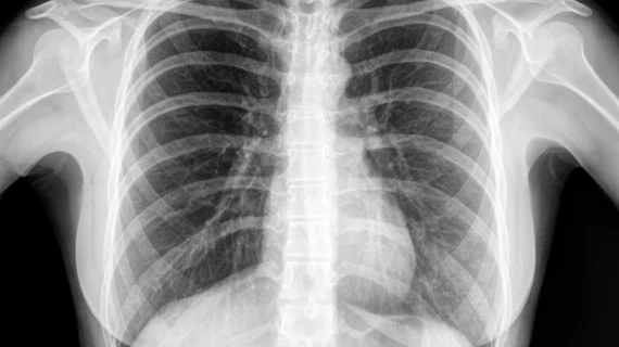
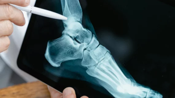
Medical X-rays have mostly converted from film, to computed radiography (CR) that used individual plates to record each X-ray digitally that then had to be uploaded into a PACS, to what is the standard-of-care today with digital radiography (DR). DR X-ray which allows immediate digital transfer of images into a PACS for immediate review. X-rays are used to diagnose fractures, bone abnormalities, lung pathologies and tumors, as well as monitor pediatric growth, plan for surgery and treat oncology patients during radiation therapy. More detailed anatomical imaging, especially soft tissue imaging, is usually sent for advanced imaging with CT or MRI. X-ray, especially mobile DR systems, are a primary use case for artificial intelligence (AI) integration. 

Obese patients undergoing x-rays have a higher cancer risk, but risk still low

The higher dose of radiation required to x-ray extremely obese patients increases those patients’ risk of cancer, according to new research published in the Journal of Radiological Protection. The chances of getting cancer due to undergoing an x-ray are still quite low, the authors added.

December 21, 2018
update policy

FDA updates diagnostic x-ray system regulations with new draft guidance

A new draft guidance from the FDA aims to clarify the agency’s policies concerning the production of diagnostic x-ray systems. The guidance, “Clarification of Radiation Control Regulations for Manufacturers of Diagnostic X-Ray Equipment,” once finalized, would replace the FDA’s previous guidance on this issue from 1989.

December 18, 2018
Project Gallery X-ray Art

New museum puts x-ray artwork on display

Artwork made out of x-ray mages is now on display at Process Gallery in Kent, a new museum located in southern England.

November 27, 2018
chestxray.jpg

Chest x-ray algorithm much faster than experienced radiologists, but not always better

Stanford University researchers have produced a convolutional neural network capable of finding most of 14 different disease states—nearly everything from pneumonia to lung cancer—on chest x-rays.

November 21, 2018

Konica Minolta Brings Motion to X-ray with Dynamic Digital Radiography at RSNA 2018

Wayne, NJ—For the first time, radiologists will be able to view motion from standard X-ray images without fluoroscopy. Konica Minolta Healthcare is bringing digital radiography (DR) to life with the ability to visualize movement using conventional X-ray. Known as Dynamic Digital Radiography (DDR)* or X-ray in Motion™, this revolutionary new modality captures movement in a single exam and allows the clinician to observe the dynamic interaction of anatomical structures, such as soft tissue and bone, with physiological changes over time. The value of DDR in thoracic imaging is promising, allowing clinicians to observe chest wall, heart and lung motion during respiration. DDR goes beyond pulmonary function; Konica Minolta is exploring its use in orthopedic applications of the spine and extremities. This new capability will be showcased at the annual meeting of the Radiological Society of North America (RSNA), being held November 25-29 in Chicago, in Konica Minolta’s booth 1919.

November 15, 2018

When training AI to classify chest x-rays, is more data always better?

Convolutional neural networks (CNNs) trained with 20,000 labeled images can accurately classify chest x-rays as normal or abnormal, according to new findings published in Radiology. Training the CNN with an additional 180,000 images, the authors noted, only yielded “marginal” benefits.

November 13, 2018

Breakthrough could lead to safer, more affordable x-ray imaging

The work of chemists from the National University of Singapore (NUS) could someday lead to safer, more affordable x-rays, according to new research published in Nature.

October 25, 2018

Patents awarded for new nanoparticle activated by x-ray, ultrasound to kill cancer

The University of Texas at Arlington (UTA) was awarded U.S. and international patents on a nanoparticle that can be activated by x-ray or ultrasound and kill multiple cancers without harming healthy surrounding tissues.

October 17, 2018

Around the web

"This was an unneeded burden, which was solely adding to the administrative hassles of medicine," said American Society of Nuclear Cardiology President Larry Phillips.

SCAI and four other major healthcare organizations signed a joint letter in support of intravascular ultrasound. 

The newly approved AI models are designed to improve the detection of pulmonary embolisms and strokes in patients who undergo CT scans.

Trimed Popup
Trimed Popup