
Portable ultrasound helps novice users accurately perform breast exams
Researchers have developed portable ultrasound-based detectors that they believe can expand access to breast imaging.
The technology comes equipped with a user interface that helps guide operators during exams. This is a valuable feature, as the quality of ultrasound exams often heavily relies on the operator and is subject to variability. Experts from MIT, where the technology was developed, are optimistic their system can be used by almost anyone, including individuals with no ultrasound experience.
The group detailed their work recently in Nature Communications.
“At each time interval, the computer interface guides you to position the device in exactly the same location, which is important for the longitudinal monitoring of a given tissue. It’s very intuitive and quite easy to use,” senior study author Canan Dagdeviren, PhD, an associate professor of media arts and sciences at MIT, said in a news release.
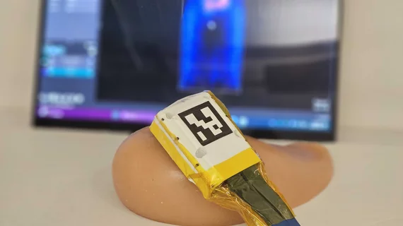
The compact system is a little larger than a smartphone. It includes an acquisition and processing module attached to a small ultrasound probe. A recent update to the probe added a “backing layer” to the transducer, which helps contain and focus the ultrasound waves. This creates more detailed images with higher resolution. The addition of an algorithm that compensate for differences in tissue types further enhances the image quality.
“With the backing layer, the device produces more accurate and sharper images, with a wider operating range of frequencies,” said Osman Goni Nayeem, the study's lead author and a former MIT postdoc MD.
The team recruited 10 volunteers with no ultrasound experience to test the updated device on a gel-like phantom. Each participant was tasked with identifying small spheres within the phantom using both traditional ultrasound equipment and the updated portable system. They yielded much stronger performances when using the system developed by MIT researchers.
Another experiment with participants imaging themselves further validated its usability. For that evaluation, seven participants were tasked with accurately placing the probe on themselves for a series of images, which they were able to do with the user interface guidance.
“Conventionally, you need an operator to move the probe around the breast, but we made a computer-vision interface for users to do it by themselves. This is very user-friendly and it shows live images on the screen,” Nayeem added.
The system could be especially beneficial in patients who have a history of breast cancer and may need additional interval exams between screenings or in those who are undergoing breast cancer treatment and need to monitor progression. In areas where skilled sonographers may be limited, the system could be used by novice users to complete these monitoring exams.
Although breast cancer is the primary focus of the team’s research, they are planning to expand their studies to other clinical scenarios in the future.
“The technology is so versatile that it can be used for any soft tissue imaging, from ovarian cancer to measuring endometriosis progression, or fetal monitoring,” Dagdeviren said.
Read more here.

In addition to her background in journalism, Hannah also has patient-facing experience in clinical settings, having spent more than 12 years working as a registered rad tech. She began covering the medical imaging industry for Innovate Healthcare in 2021.