
AI model helps discern patients' need for supplemental breast imaging
A novel deep learning model could offer providers greater insight into the need for supplemental breast imaging than current risk criteria, according to new research.
Published in JAMA Network Open, findings suggest that the model is more accurate than current risk assessments that are based on breast density when it comes to determining whether patients need additional imaging beyond breast cancer screening. When tasked with providing a five-year cancer risk assessment for over 120,000 screening mammograms, the model was significantly more accurate than density category assignments, suggesting that it could offer a more precise alternative.
Authors of the paper believe AI has the potential to serve as an alternative to previous risk criteria.
“Providing access to supplemental imaging for all women with dense breasts, regardless of their actual risk, may result in overuse of limited imaging resources, increase false-positive results, and impose financial burdens, particularly in underserved populations,” Leslie R. Lamb, MD, with the department of radiology at Massachusetts General Hospital, and colleagues explained. “To achieve more personalized and equitable screening, there is a need for more precise risk stratification tools that go beyond breast density alone.”
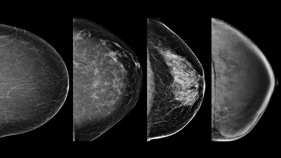
The model (Mirai, Massachusetts Institute of Technology and Mass General) was designed to calculate a woman’s five-year risk based on subtle patterns on screening mammograms; it categorizes risk as low (<1.7%), intermediate (1.7%–3%) or high (>3%). Researchers applied it to around 123,000 screening mammograms acquired across five sites at a large academic health system between 2009 and 2018. The model’s risk assessments were compared to the exams’ BI-RADS categories and follow-up data using cancer diagnoses and false negatives to gauge its prediction accuracy.
Around 41% of the exams included in the analysis were classified as dense. The team observed a significant improvement (AUROC of 0.71 vs. 0.53) for predicting five-year risk via the model in comparison to density classifications alone. However, it did not yield greater discriminatory potential with respect to false negatives; the group noted an increase in false negatives across the model’s risk groups, especially among women with dense breasts (2.1 per 1,000 examinations in high-risk vs. 1 and 0.6 in intermediate and low-risk groups). Integrating density status into the model’s predictions did not yield an improvement.
“Importantly, DL risk scores remained predictive in women with nondense breasts, suggesting that current binary density-based policies may both under-identify high-risk women without dense breasts and over-identify low-risk women with dense breasts for supplemental imaging,” the authors noted. “The absence of performance improvement with the addition of breast density supports the assumption that density-related imaging features are already incorporated within the DL model’s risk estimates.”
The group suggested their findings support the routine use of DL-based risk assessments, as this method can reduce unnecessary imaging and improve diagnostic accuracy.
“In the current era of density legislation, DL models could also be used to identify the subset of women with dense breasts most likely to benefit from supplemental imaging while sparing low-risk women unnecessary imaging,” they added.”
Read more here.

In addition to her background in journalism, Hannah also has patient-facing experience in clinical settings, having spent more than 12 years working as a registered rad tech. She began covering the medical imaging industry for Innovate Healthcare in 2021.