Breast Imaging

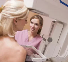
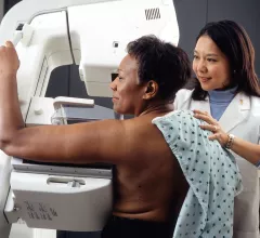
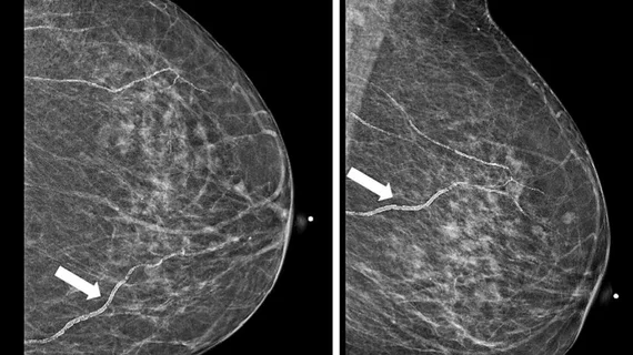
Breast imaging includes imaging modalities used for breast cancer screenings and planning therapy once cancer is detected. Mammography is the primary modality used. Mammogram technology is moving from 2D full-field digital mammography (FFDM) to breast tomosynthesis, or 3D mammography, which helps reduce false positive exams by allowing radiologists to look through the layers of tissue. Overlapping areas of dense breast tissue on 2D mammograms appear similar to cancers and 3D tomo helps determine if suspect areas are cancer or not. About 50% of women have dense breast tissue, which appears white on mammograms, the same as cancers, making diagnosis difficult. Radiologists use the Breast Imaging Reporting and Data System (BI-RADS) scoring system to define the density of breast tissue. Many states now require patients to be notified if they have dense breasts so they understand their mammograms might be suboptimal and they should use supplemental imaging that can see through the dense areas. This includes tomosythesis, breast ultrasound, automated breast ultrasound (ABUS), breast MRI, contrast enhanced mammography and nuclear imaging, including positron emission mammography (PEM).
Thumbnail

There’s no stopping 3D mammography now, but can it sustain this pace?

Seven years after the FDA approved the first tomosynthesis device for breast cancer screening, adoption rates for digital breast tomosynthesis (DBT) remain on an upswing. Earlier this year the agency reported a nearly 30 percent increase of certified mammography facilities offering DBT—aka “3D mammography,” aka “tomo”—over just the past year (from 3,178 facilities in March 2017 to 4,074 in March 2018).

Thumbnail

ACR, SBI publish new breast cancer screening guidelines

The American College of Radiology (ACR) and Society of Breast Imaging (SBI) have published new breast cancer screening guidelines, once again recommending that all women begin annual screening at age 40.

2018 SBI/ACR Breast Imaging Symposium headed to Las Vegas

The Society of Breast Imaging (SBI) is working with the American College of Radiology (ACR) to host its annual Breast Imaging Symposium April 12-15 in Las Vegas.

Thumbnail

Q&A: Jay A. Baker on breast cancer screening, the benefits of DBT and more

Jay A. Baker, MD, professor of radiology and chief of the breast imaging division at Duke University Medical Center in Durham, North Carolina, and the vice president of the Society of Breast Imaging (SBI), spoke with Radiology Business about some of the biggest topics affecting breast imaging today.

Pivotal Trial Data Support Potential of Seno Medical Instruments' Imagio™ Opto-Acoustic Breast Imaging System in Reducing False Positive Breast Examinations and Benign Biopsies

CHICAGO, Nov. 27, 2017 /PRNewswire/ -- Seno Medical Instruments, Inc. (Seno Medical), the leader in new technology for breast cancer diagnosis using opto-acoustic ultrasound (OA/US) imaging to differentiate benign from malignant masses, today announced positive data from PIONEER, a Phase III pivotal trial of its Imagio™ breast imaging system. The study found that OA/US was more specific than device gray-scale ultrasound alone (US) in differentiating malignant from benign breast lesions and was non-inferior to US with respect to sensitivity.

Thumbnail

Breast and Lung Cancer Screening: The Push for Patient Engagement

Shortly before her 40th birthday, a woman visits her primary care physician and is advised she should schedule her first mammogram. The patient is puzzled and says she “read somewhere” that she can wait a few years. The physician has heard this before and kindly convinces the patient to comply with the recommendation. She agrees, schedules the mammogram and receives her results before even leaving the imaging facility. 

Thumbnail

Mammography disparities found among black, Hispanic women

While early screening and detection can play an important role in preventing breast cancer, new research from Mayo Clinic shows that some women, particularly minority women, aren’t being screened as often as their white counterparts. 

Example of a mammogram showing X-ray images of both the right and left breast and patches of dense breast tissue.

Healthcare groups push for extension of mandate that guarantees coverage of annual mammograms

Multiple patient advocacy groups and healthcare organizations, including the American College of Radiology (ACR) and Society of Breast Imaging (SBI), are asking Congress to extend a federal mandate that requires insurance companies to fully insure annual mammograms for women 40 years old and older. 

Around the web

The nuclear imaging isotope shortage of molybdenum-99 may be over now that the sidelined reactor is restarting. ASNC's president says PET and new SPECT technologies helped cardiac imaging labs better weather the storm.

CMS has more than doubled the CCTA payment rate from $175 to $357.13. The move, expected to have a significant impact on the utilization of cardiac CT, received immediate praise from imaging specialists.

The all-in-one Omni Legend PET/CT scanner is now being manufactured in a new production facility in Waukesha, Wisconsin.

Trimed Popup
Trimed Popup