Medical Imaging

Physicians utilize medical imaging to see inside the body to diagnose and treat patients. This includes computed tomography (CT), magnetic resonance imaging (MRI), X-ray, ultrasound, fluoroscopy, angiography,  and the nuclear imaging modalities of PET and SPECT. 

MRI scans using diffusion tensor imaging can predict Alzheimer's disease

MRI scans using diffusion tensor imaging (DTI) show potential for predicting if a patient will develop Alzheimer’s disease later in life, according to findings to be presented at RSNA 2018 in Chicago.

Thumbnail

AI startup Ezra secures $4M for MRI-based prostate cancer screening program

Ezra, a New York City-based artificial intelligence (AI) startup, has secured $4 million in funding for its new direct-to-consumer prostate cancer screening program.

Thumbnail

MRI captures more detail in colon cancers than multidetector CT

MRI may be a better choice than multidetector CT for identifying high-risk colon cancers that have already reached stage II or stage III, are still surgically resectable and are at risk of progressing to stage IV.

Thumbnail

'Brightness' on ultrasound images helps radiologists predict type 2 diabetes

When ultrasound reveals shoulder muscles that appear especially “bright,” it may be a warning sign of diabetes, according to findings to be presented at RSNA 2018 in Chicago.

Thumbnail

MR spectroscopy adds little diagnostic value when imaging brain tumors

Adding magnetic resonance spectroscopy (MRS) to MRI does not significantly improve the classification of brain tumors in clinical practice, although MRS may be a valuable supplement to MRI in certain cases, according to researchers from Sweden's Uppsala University.

Konica Minolta Brings Motion to X-ray with Dynamic Digital Radiography at RSNA 2018

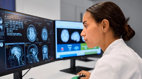
Wayne, NJ—For the first time, radiologists will be able to view motion from standard X-ray images without fluoroscopy. Konica Minolta Healthcare is bringing digital radiography (DR) to life with the ability to visualize movement using conventional X-ray. Known as Dynamic Digital Radiography (DDR)* or X-ray in Motion™, this revolutionary new modality captures movement in a single exam and allows the clinician to observe the dynamic interaction of anatomical structures, such as soft tissue and bone, with physiological changes over time. The value of DDR in thoracic imaging is promising, allowing clinicians to observe chest wall, heart and lung motion during respiration. DDR goes beyond pulmonary function; Konica Minolta is exploring its use in orthopedic applications of the spine and extremities. This new capability will be showcased at the annual meeting of the Radiological Society of North America (RSNA), being held November 25-29 in Chicago, in Konica Minolta’s booth 1919.

Abdominal ultrasound often not enough for children with suspected appendicitis

Children presenting with signs of appendicitis should receive careful further evaluation if the usual first-line exam, abdominal ultrasound, fails to deliver a clear view of the vestigial digestive-system organ.

Thumbnail

Pipe organ-like ultrasound transducer offers improved quality for medical images

Researchers from the University of Strathclyde in Glasgow, Scotland, have developed a miniaturized version of a musical pipe organ, or a piezoelectric micromachined ultrasonic transducer (PMUT), that could potentially improve the quality of medical images.

Around the web

The ACR hopes these changes, including the addition of diagnostic performance feedback, will help reduce the number of patients with incidental nodules lost to follow-up each year.

And it can do so with almost 100% accuracy as a first reader, according to a new large-scale analysis.

The patient, who was being cared for in the ICU, was not accompanied or monitored by nursing staff during his exam, despite being sedated.