Can iterative metal artifact reduction improve CT image quality in gunshot victims?
Though its effects vary based on presets and tissue densities, iterative metal artifact reduction (iMAR) could present a unique opportunity to image gun trauma patients whose scans are otherwise compromised by artifacts from retained bullets, researchers wrote in the European Journal of Radiology this month.
Radiologic technologist Dominic Gascho and colleagues led a study that examined the use of iMAR in a phantom setting and a porcine setting, using a severed pig’s head to mimic human tissue. The authors said that with a global increase of violence comes an increase of patients with gunshot wounds. Postprocessing methods like frequency-split metal artifact reduction, normalized metal artifact reduction and monoenergetic dual-energy CT have shown only limited promise.
“Each of these methods imposed different challenges in reducing metallic artifacts due to differences in the implant composition and local anatomy,” Gascho et al. wrote in EJR. “A recently introduced software algorithm for iMAR has been demonstrated to significantly reduce artifacts by combining a frequency-split technique with normalized sinogram interpolation.”
iMAR allows for the selection of different reconstruction presets for a host of familiar medical implants, like neuro-coils, dental fillings, pacemakers and hip implants. Those presets allow for more painless reconstruction of images that may have been obstructed by bullet fragments, the authors said.
“Metallic hardware frequently results in considerable streaking artifacts on CT due to photon starvation and beam-hardening,” they wrote. “These artifacts degrade the image quality and may obscure critical anatomical structures or pathological findings, especially in the region near the metallic foreign body.”
That means that in the worst case scenario, retained bullets conceal relevant pathologies, making for non-diagnostic images in gunshot patients. Reconstruction itself could be difficult, too, Gascho and co-authors said, because those fragments could interfere with the true bullet path.
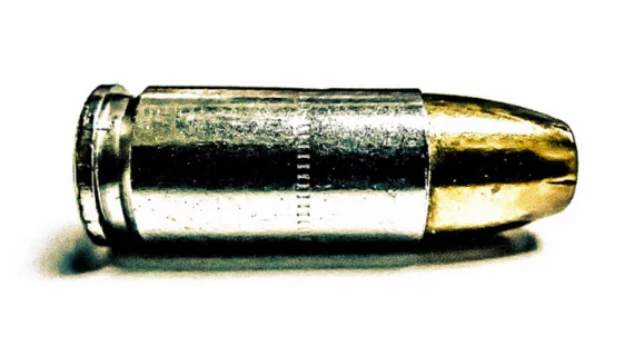
For their study, Gascho and his team selected nine different types of projectiles to evaluate the efficacy of iMAR. Each projectile was fixed on a thin thread and placed in a container of water to reflect a homogeneous medium, then placed in the pig’s head as a substitute for human tissue. Raw data from all CT scans were reconstructed with and without iMAR, and eventual reconstructions were evaluated by five radiologists and five residents.
The researchers found that in the phantom setting, iMAR presets for neuro-coils and extremity implants were most highly rated, while in the pig’s head setting with the soft tissue window, neuro-coils and extremity implant presets were preferred.
“The applied iMAR presets revealed different effects on the image quality,” the authors wrote. “The results of this study indicate that the neuro-coils preset is the most appropriate preset for soft tissue, and the preset extremity implants is favorable for bones in cases of retained bullets.”

