Phase-contrast CT helps researchers see Egyptian mummy up close and personal
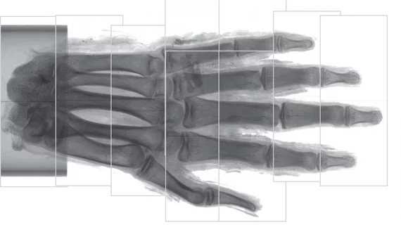
Researchers have imaged the soft tissue of an Egyptian mummy’s hand using phase-contrast CT, according to research published in Radiology. What does this mean for medical researchers going forward?
Though x-ray and CT have been used in the past for imaging ancient remains through absorption-contrast imaging, phase-contrast CT can help specialists capture even better, more detailed images.
“There is a risk of missing traces of diseases only preserved within the soft tissue if only absorption-contrast imaging is used,” co-author Jenny Romell, MSc, of KTH Royal Institute of Technology/Albanova University Center in Stockholm, Sweden, said in a prepared statement. “With phase-contrast imaging, however, the soft tissue structures can be imaged down to cellular resolution, which opens up the opportunity for detailed analysis of the soft tissues.”
The hand, from the Museum of Mediterranean and Near Eastern Antiquities, was brought to Sweden back in the 19th century. It is believed to be from around 400 BCE (before common era). The resolution of the final images was “between 6 to 9 micrometers, or slightly more than the width of a human red blood cell,” according to the statement.
“Just as conventional CT has become a standard procedure in the investigation of mummies and other ancient remains, we see phase-contrast CT as a natural complement to the existing methods,” Romell said. “We hope that phase-contrast CT will find its way to the medical researchers and archaeologists who have long struggled to retrieve information from soft tissues, and that a widespread use of the phase-contrast method will lead to new discoveries in the field of paleopathology.”

