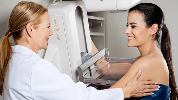
A key difference between synthetic mammography, FFDM
Measured mass sizes are considerably smaller on synthetic mammography (SM) images than full-field digital mammography (FFDM) images, according to new findings published in Academic Radiology.
“Ideally, mass size measurements are not expected to change between FFDM and SM images,” wrote Halit Nahit Şendur, MD, Gazi University Faculty of Medicine in Turkey, and colleagues. “However, there are limited numbers of studies comparing lesion sizes between SM and FFDM in the literature. Therefore, we aimed to determine whether SM technology had an effect on mass size measurements and compared interobserver variability in mass size measurements between SM and FFDM images.”
Şendur et al. explored data from 143 patients who underwent both FFDM and digital breast tomosynthesis (DBT) from June 2018 to February 2019. SM images were reconstructed using the DBT data. Two observers—one with one year of experience and another with four years of experience—measured mass sizes independently at two different times two weeks apart. During the first session, the readers only looked at the FFDM images. The second session only included SM images.
Overall, the authors found that mass size measurements were “significantly smaller” on SM. Also, the interobserver differences were greater than the differences between FFDM and SM images.
The mean mass size for FFDM images was 20.27 mm ± 14.10 for the first observer and 21.56 ± 14.84 for the second observer. The mean mass size for SM images was 18.50 mm ± 13.05 for the first observer and 19.89 ± 13.68 mm for the second observer.
“These findings suggested that the algorithm used to create SM images had an impact on the dimensions of lesions,” the authors wrote. “Therefore, our results may provide a guide for the specific aspects of the SM software that should be improved.”
It is also possible, the authors explained, that these results could “illustrate an additional advantage of SM technology.”
“Because the signal obtained from DBT is ideally reconstructed into the correct plane above the detector, viewing masses in DBT slices, and likely on SM images, may lead to measure masses at truer sizes,” they wrote.
Şendur and colleagues did note that their work had certain limitations, including the fact that the actual mass sizes were not a part of the study.
“In the current study, we aimed to document discrepancy of mass size measurements between FFDM and SM images rather than assessing which imaging technique provides more accurate measurements,” the authors wrote. “As the comparison of accuracy of mass size measurements between these two imaging techniques was not in the scope of this study, histological sizes were not required. Future studies which include histological size measurements and comparing the accuracy of SM and FFDM images will be valuable.”
Another limitation was that SM images were all created using the same vendor’s algorithm. Using DBT solutions from a variety of vendors could help researchers learn even more about SM.

