
Portable system captures 1st diagnostic-quality X-rays during spaceflight
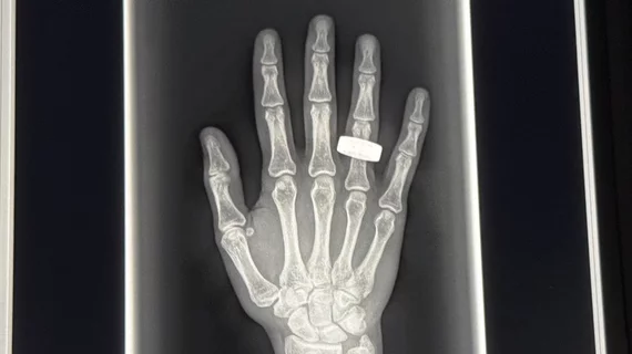
The ability to capture diagnostic radiographs in-orbit may soon be routine during spaceflight.
Ultrasound imaging has been available during space missions for years. However, these exams are challenging to acquire and are highly dependent on operator skill, making their quality variable and subject to misinterpretation by readers. Now, a new paper published in RSNA’s flagship journal Radiology details how experts were able to successfully capture diagnostic radiographs using an off-the-shelf portable X-ray system. Acquired during a 3.5-day polar orbital flight on SpaceX’s Fram2 in 2025, the radiographs were captured by crew members who underwent just four hours of training prior to the mission.
This feat was previously perceived as “too technically challenging,” but this latest success has experts hopeful that they are now one step closer to being able to provide diagnostic images beyond ultrasound alone during spaceflight.
“Portable X-ray machines are in use everywhere—at the Kentucky Derby, on the sidelines of the Super Bowl and around the globe in low-resource areas—because they can run on solar power and can be operated by individuals with no medical expertise,” Sheyna Gifford, MD, lead researcher and an assistant professor of aerospace medicine at Mayo Clinic in Rochester, Minnesota, said in a news release from the Radiological Society of North America. “We believed an off-the-shelf portable system would stand a very good chance of surviving prelaunch testing and be operational in space by crew members with minimal training.”
The X-rays were acquired using an FDA-cleared, ultraportable system and flat panel detector developed by MinXray, KA Imaging and prominent academic institutions. After training, three participating crew members captured preflight and in-flight images of a hand, forearm, abdomen, pelvis and chest without remote assistance. Images of equipment also were acquired. The images were evaluated by a group of three independent radiologists for overall image quality, spatial resolution, contrast resolution and positioning.
The in-flight and preflight radiographs of the extremities were similar in terms of quality, spatial and contrast resolution. Images of the chest, abdomen and pelvis, however, differed in positioning quality, with the in-flight images slightly inferior. Images of equipment enabled visualization of internal equipment components to the submillimeter scale. Despite the equipment sustaining some damage on re-entry, it continued to function as expected upon landing.
These results highlight the promise of portable imaging in the future of spaceflight, Gifford suggested.
“By acquiring the first human and equipment X-rays in space, our study demonstrates the feasibility of in-orbit radiography and expanded diagnostic capabilities for crew health and hardware evaluation,” Gifford said. “Acquiring a diagnostically useful X-ray in space is something that anyone can do. Three very talented nonmedical people with four hours of training in one of the harshest environments did it right and did it well.”
Read more here.

In addition to her background in journalism, Hannah also has patient-facing experience in clinical settings, having spent more than 12 years working as a registered rad tech. She began covering the medical imaging industry for Innovate Healthcare in 2021.