
Body composition changes on CT could signal pancreatic cancer years before diagnosis
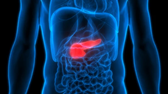
Artificial intelligence-enabled analyses of routine CT scans could help identify changes in body fat linked to pancreatic cancer years before the disease is diagnosed.
That’s according to new research out of Mayo Clinic. Published in Pancreatology, the study indicates that imaging-derived body composition measurements could help identify patients at increased risk of pancreatic cancer, enabling provides to initiate treatment before the disease has progressed.
The study examined body composition changes in patients who later developed pancreatic ductal adenocarcinoma, the most common form of pancreatic cancer. Unfortunately, the disease is frequently diagnosed at an advanced stage, and most cases occur in people who do not meet current criteria for high-risk surveillance, which allows the cancer to progress before it is diagnosed.
“Pancreatic cancer often develops silently, making early detection extremely challenging,” Yan Bi, MD, PhD, a gastroenterologist at Mayo Clinic and senior author of the study, said in a news release on the results. “These findings suggest that routine CT scans may capture physiological changes associated with pancreatic cancer before patients are diagnosed.”
For the study, researchers analyzed abdominal CT scans from 1,825 patients with pancreatic ductal adenocarcinoma and 3,650 matched controls across Mayo Clinic sites in Arizona, Florida and Minnesota. Using an AI-based body composition tool, they measured changes in skeletal muscle, visceral fat and subcutaneous fat over time.
During the three years before diagnosis, patients who eventually developed pancreatic cancer showed significantly steeper declines in both visceral and subcutaneous fat than matched controls. Differences in muscle mass were most apparent during the final 12 months before diagnosis.
Mayo researchers also assessed whether changes in body composition could help distinguish patients who later developed cancer from those who did not. For this, the measurements showed moderate discriminatory ability. This, the research group suggested, indicates that longitudinal changes in fat and muscle could contribute to risk assessment approaches in the future.
The results highlight a potential role for opportunistic analysis of imaging already obtained for other clinical reasons via automated body composition measurements. This could help identify patterns that might otherwise be overlooked during routine clinical care.
Read more here.

In addition to her background in journalism, Hannah also has patient-facing experience in clinical settings, having spent more than 12 years working as a registered rad tech. She began covering the medical imaging industry for Innovate Healthcare in 2021.