
Visible breast lesions often overlooked on emergency chest CT scans
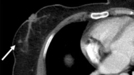
A significant number of incidental breast masses are overlooked on CT scans of the chest, which could potentially lead to delays in diagnosis and treatment, researchers caution.
A new Academic Radiology analysis of incidentally detected cancers suggests that up to two-thirds of visible breast masses on emergency chest CTs go unnoticed by readers. At once center, such misses resulted in significant delays in cancer diagnosis, up to 33 months in some cases.
Although emergency chest CT scans are usually completed for other clinical indications, experts involved in the study say they present an opportunity to catch cancer earlier. However, visible lesions on these scans often are overlooked.
“In recent years, especially with the COVID-19 pandemic, a significant increase in CT use in emergency departments has been observed,” Olgun Sahin, MD, with the Department of Radiology, Izmir City Hospital in Turkey, and colleagues explained. “The increasing number of chest CT scans is assumed to lead to a corresponding increase in the detection of incidental breast cancers. However, despite this increased detection potential, published data on clinical practice is striking. A retrospective study by Park et al. revealed that 64.3% of incidental breast cancers were missed in initial chest CT reporting. Similarly, Abougazia et al. recently reported 67.9% of such cases.”
Researchers sought to determine whether lesion- or patient-specific factors played a role in visible cancers being missed on emergency chest CT scans. They analyzed the cases of 64 patients who presented to the ED with non-breast-related complaints. Each had either BI-RADS 4 or BI-RADS 5 lesions with histopathological diagnoses of malignancy in the months or years after their CT scans. Lesions were classified as “detected” or “missed,” with researchers taking note of specific details of each to identify features that were more likely to lead to a lesion being overlooked.
Of the 73 scans, a discrete breast mass or a focal asymmetric breast tissue density was detected and reported in around 30%; conversely, 70% were missed, leading to delays in cancer diagnoses ranging from 4 to 33 months. Missed lesions were typically smaller, with the mean size being 13.6 mm compared to 22.2 mm for detected lesions. Missed lesions were more commonly located in the right breast, while detected lesions were more often in the inner zone and accompanied by axillary lymphadenopathy and a fatty (Type A) breast parenchymal pattern.
Given the healthcare environment where these scans were completed, the team suggested that the areas where the lesions were located were likely not the radiologist's focus. Systematic evaluation of breast parenchyma and inclusion of breast density in CT reports may help remedy the issue, as could artificial intelligence algorithms designed to spot incidental findings.
“AI-assisted image analysis systems may serve as a valuable adjunct in reducing missed incidental breast lesions on chest CT examinations,” the authors noted. “These systems have significant potential in identifying lesions that might be overlooked by radiologists, particularly those that are small in size or morphologically atypical, thereby substantially enhancing diagnostic sensitivity.”
Read more here.

In addition to her background in journalism, Hannah also has patient-facing experience in clinical settings, having spent more than 12 years working as a registered rad tech. She began covering the medical imaging industry for Innovate Healthcare in 2021.