Computed Tomography

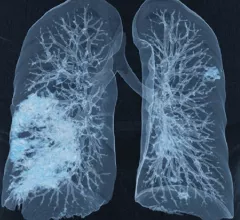
Computed tomography (CT) is a fast and accurate imaging modality often used in emergency settings and trauma imaging. CT scans, with or without (or both) iodinated contrast are frequently used to image the brain, chest, abdomen and pelvis, but also have post-imaging reconstructive capabilities for detailed orthopedic imaging. It is now a standard imaging modality in emergency rooms to quickly assess patients. CT uses a series of X-ray images shot as the gantry rotates around the patient. Computer technology assembles these into into a dataset volume than can be slices on any access, or advanced visualization software can extract specific parts of the anatomy for study. Find more content specific to cardiac CT.


Thumbnail

MRI catches CT in head-to-head lung imaging

When it comes to assessing patients with suspected pulmonary embolism, contrast-enhanced CT pulmonary angiography has no diagnostic edge over a certain free-breathing, unenhanced MRI perfusion protocol.

Thumbnail

Including radiologists in radiotherapy prep bolsters quality of care

Many radiation oncologists are not formerly trained in imaging interpretation, and radiologists’ collaborative participation in care planning can help to catch errors, experts wrote recently.

Ed Nicol, MD, consultant cardiologist and honorary senior clinical lecturer with Kings College London and president-elect of the Society of Cardiovascular Computed Tomography (SCCT), explained artificial intelligence (AI) in cardiac CT is here to stay and its use is expanding. He noted that one AI-based algorithm is already included in recent cardiology guidelines and more will likely follow. #SCCT

Cardiac imagers need to understand AI as it enters clinical use and ACC guidelines

Most FDA-cleared AI algorithms are related to radiology and cardiology, meaning radiologists and cardiologists need to make an effort to learn how these technologies work.

Internal surgery patients with infectious complications shown not to benefit by extra CT

Thoracic CT is safely and wisely omitted from diagnostic protocols for ICU patients who have signs of infection after abdominopelvic surgery but were already imaged with abdominal CT.

Homegrown tracking boosts follow-up imaging for incidental findings of uncertain gravity

A structured program to track incidental findings on body CT has significantly boosted rates of clinician follow-up as well as timely patient adherence to radiologist recommendations for next exams.

Appearances can be deceiving on chest CT performed for COVID in cancer patients

In a study of more than 250 COVID-positive patients with a history of any cancer, fewer than half the cohort had chest CT findings deemed typical for COVID-related pneumonia based on an RSNA classification guide. 

Q4 and full-year 2022 results in for GE HealthCare

GE HealthCare has some favorable tailwinds at its back as it proceeds into its first year unattached to a certain venerable parent corporation. 

AI helps reading-room radiologists differentiate colon cancer from diverticulitis

The model augmented and significantly improved diagnostic performance for abdominal subspecialists as well as residents—a result researchers say has major clinical implications.

Around the web

The all-in-one Omni Legend PET/CT scanner is now being manufactured in a new production facility in Waukesha, Wisconsin.

After reviewing years of data from its clinic, one institution discovered that issues with implant data integrity frequently put patients at risk. 

Prior to the final proposal’s release, the American College of Radiology reached out to CMS to offer its recommendations on payment rates for five out of the six the new codes.

Trimed Popup
Trimed Popup