Computed Tomography

Computed tomography (CT) is a fast and accurate imaging modality often used in emergency settings and trauma imaging. CT scans, with or without (or both) iodinated contrast are frequently used to image the brain, chest, abdomen and pelvis, but also have post-imaging reconstructive capabilities for detailed orthopedic imaging. It is now a standard imaging modality in emergency rooms to quickly assess patients. CT uses a series of X-ray images shot as the gantry rotates around the patient. Computer technology assembles these into into a dataset volume than can be slices on any access, or advanced visualization software can extract specific parts of the anatomy for study. Find more content specific to cardiac CT.


M&A mergers and acquisitions business deal

GE HealthCare eyes IR guidance company, releases revenue results

Less than a week after ringing the Nasdaq opening bell to showcase its long-awaited independence, GE HealthCare has announced a first strategic acquisition is in the works.

Mahadevappa Mahesh, PhD, professor of radiology and a medical physicist at the Johns Hopkins University School of Medicine, explains a new American College of Radiology (ACR) effort to ensure that lower radiation dose X-ray images under Image Wisely and As Low as Reasonable Achievable (ALARA) meet diagnostic reading standards. He spoke to Radiology Business at the Radiological Society of North America (RSNA) 2022 meeting.

VIDEO: Making sure lower dose X-ray is still diagnostic quality

Mahadevappa Mahesh, PhD, professor of radiology and a medical physicist at the Johns Hopkins University School of Medicine, explains a new ACR effort to ensure that lower radiation dose X-ray images under Image Wisely and As Low as Reasonable Achievable (ALARA) meet diagnostic reading standards.

A CT coronary calcium scoring exam at Northwestern Medicine's Central DuPage Hospital in the Chicago Suburbs. Dave Fornell

Trends in the number of radiology studies and a decline in radiation dose

A special report in Radiology offers a rare overview of the number of radiology exams performed each year worldwide and focuses on the decline of associated radiation doses patients receive.

Urinary stones in the ED: What will it take for ultrasound to gain ground on costly, radiative CT?

Professional consensus supports the use of ultrasound for initial imaging evaluation of patients presenting in the ED with suspected urinary stone disease (USD). However, as of 2018, only 2% of these patients received ultrasound while some 59% had CT.

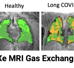
Sean Fain, PhD, vice chair of radiology and research and a professor of radiology, Division of Cardiovascular and Thoracic Imaging, University of Iowa, discusses how long-COVID lung damage can be tracked using xenon (Xe) gas magnetic resonance imaging (MRI) and quantitative computed tomography (CT). He spoke to Health Imaging at RSNA 2022.

VIDEO: Tracking long-COVID lung damage using MRI and CT

Sean Fain, PhD, vice chair of radiology and research and a professor of radiology, University of Iowa, discusses how long-COVID lung damage can be tracked using xenon (Xe) gas MRI and quantitative CT at RSNA 2022. 

Imaging industry names in the news: Koning, Medality, QT Imaging, Rezolut, Scanslated, more

Noteworthy market developments listed in the order announcements were posted.    

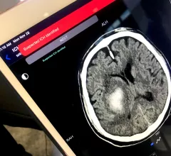
An example of artificial intelligence (AI) automated detection of a intracranial hemorrhage (ICH) in. a CT scan used to send alerts to the stroke acute care team before a radiologist even sees the exam. Example shown by TeraRecon at RSNA 2022.

VIDEO: Radiology AI aids acute care and other departments

Sanjay Parekh, PhD, senior market analyst with Signify Research, explains how some radiology AI is being adopted outside of radiology departments to improve care.

A stack deep silicon photon counting CT detector components. The University of Wisconsin is working with GE Healthcare to develop the new PCD technology. The system and the deep silicon technology was unveiled at RSNA 2022. #RSNA22 #PCCT

VIDEO: Photon-counting CT development at the University of Wisconsin

Tim Szczykutowicz, PhD, DABR, associate professor of radiology at the University of Wisconsin-Madison, is helping develop a new type of photon-counting CT detector that was shown as a work-in-progress by GE Healthcare at RSNA 2022.

Around the web

The nuclear imaging isotope shortage of molybdenum-99 may be over now that the sidelined reactor is restarting. ASNC's president says PET and new SPECT technologies helped cardiac imaging labs better weather the storm.

CMS has more than doubled the CCTA payment rate from $175 to $357.13. The move, expected to have a significant impact on the utilization of cardiac CT, received immediate praise from imaging specialists.

The all-in-one Omni Legend PET/CT scanner is now being manufactured in a new production facility in Waukesha, Wisconsin.

Trimed Popup
Trimed Popup