Computed Tomography

Computed tomography (CT) is a fast and accurate imaging modality often used in emergency settings and trauma imaging. CT scans, with or without (or both) iodinated contrast are frequently used to image the brain, chest, abdomen and pelvis, but also have post-imaging reconstructive capabilities for detailed orthopedic imaging. It is now a standard imaging modality in emergency rooms to quickly assess patients. CT uses a series of X-ray images shot as the gantry rotates around the patient. Computer technology assembles these into into a dataset volume than can be slices on any access, or advanced visualization software can extract specific parts of the anatomy for study. Find more content specific to cardiac CT.


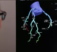
Heartflow FFR-CT allows noninvasive assessment of a patient'c coronary arteries without the need for a diagnostic angiogram.

VIDEO: HeartFlow FFR-CT sees increased interest after inclusion in the 2021 Chest Pain Guidelines

HeartFlow’s non-invasive cardiac computed tomography derived fractional flow reserve (FFR-CT) technology enables FFR values for the entire coronary tree of a patient using just a CT scan. This can show the significance of any coronary plaque lesions and if a patient needs to be sent to the cath lab for revascularization, or if they can be treated medically.

Imaging shows COVID vaccines effective at warding off pulmonary embolism

Researchers have found the condition significantly less among patients who received at least two doses of a COVID vaccine.

AI aids coma prognostics, potentially averting withdrawal of care

Deep learning has bested experienced neurosurgeons at predicting poor outcomes, including mortality, among patients admitted comatose with severe traumatic brain injuries.

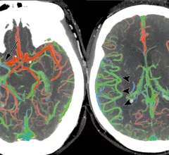
Ischemic stroke CT scan showing color coded blood flow for early and later arterial and venous contrast phases and areas of blocked blood flow. Image courtesy of RSNA

Disparities evident as CT stroke imaging rises sharply over 7-year period

Utilization of head CT perfusion (CTP) for imaging stroke patients spiked 428% between 2012 and 2019. Curiously, the uptrend was associated with higher mortality up to a year after discharge.

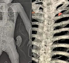
Radiology imaging of two wounded civilians with bullet and shrapnel embedded in tissue. Left is a digital X-ray of a patient with shrapnel in the chest and a bullet in the leg who was brought to the National Children's Specialized Hospital in Kyiv. Right, a 3D CT scan reconstruction of a bullet in the chest of a 40-year-old father who as attempting to evaculate his wife and child from the Russian advance near Kyiv when the Russians opened fire on him. Image from the Ukraine Heart Institute in Kyiv.

Radiologists in Ukraine helping the war effort as Russia continues to attack

Many radiologists stepped up to do their part for the war effort by staying at their hospital posts and helping the wounded as they started coming in.

Why cardiologists and radiologists are choosing cardiac CT over invasive angiography for suspected CAD

"A CT-first strategy allows physicians to provide relevant findings without the need for an invasive procedure," one radiologist explained. 

PHOTO GALLERY: Duly Health adopts outpatient cardiac CT as a standard of care

Duly Health and Care in suburban Chicago recently opened a new outpatient cardiac evaluation center equipped with a dedicated cardiac CT system, which will likely be a new business model that will be seen more in the coming years.

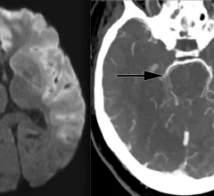
Ischemic stroke CT imaging. Images courtesy of RSNA

VA telestroke program prevents unnecessary hospital transfers and improves rural outcomes

A new study of the U.S. Department of Veterans Affairs Veterans Health Administration (VHA) National Telestroke Program shows the program prevents unnecessary hospital transfers for stroke patients and increases outcomes for patients at rural VA clinics.

Around the web

The nuclear imaging isotope shortage of molybdenum-99 may be over now that the sidelined reactor is restarting. ASNC's president says PET and new SPECT technologies helped cardiac imaging labs better weather the storm.

CMS has more than doubled the CCTA payment rate from $175 to $357.13. The move, expected to have a significant impact on the utilization of cardiac CT, received immediate praise from imaging specialists.

The all-in-one Omni Legend PET/CT scanner is now being manufactured in a new production facility in Waukesha, Wisconsin.

Trimed Popup
Trimed Popup