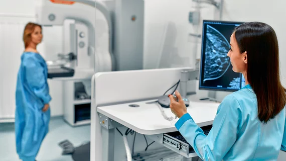
Diagnostic screening programs help catch cancer, abnormalities or other diseases before they reach an advanced stage, saving lives and healthcare costs. Screening programs include, lung, breast, prostate, and cervical cancer, among many others.
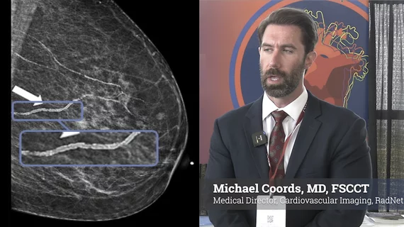
The law reflects growing interest in using existing imaging studies to identify cardiovascular disease risk without requiring additional scans, radiation exposure or costs.
New research could provide useful information for cancer registries and potentially help policymakers better evaluate screening outcomes to develop more effective guidelines.
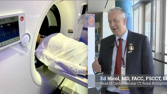
Former SCCT president Ed Nicol, MD, knows there has been interest in the potential of automated CT exams. However, he isn't so sure they are possible at this point in time for a number of reasons.
Training using transesophageal echocardiography (TEE) simulators typically relies on grainy drawings of patients in black and white. But a new version, developed by a team from the University of Washington led by Florence Sheehan, MD, can display 3D images of a hypothetical patient, allowing users to look for blood clots and other abnormalities.
A Children’s National Health System team has found velocity-selective arterial spin labeling (VSASL), an advanced MRI technology, can detect early signs of global placental perfusion in pregnancies complicated by fetal congenital heart disease (CHD).
Researchers, investigating how community practices are following breast cancer screening guidelines, found that high-risk individuals, especially those with a family history of the disease, were not receiving additional MRI scans to help in early detection.
Perhaps rules are meant to be broken. But guidelines, according to recent research, are not always followed when physicians recommend breast cancer screening to patients.
Dense breast tissue presents problems for detecting cancer—often leading to additional screenings after negative or questionable mammograms. But at RSNA 2017 in Chicago, a research team discussed abbreviated breast MRI (AB-MR), a new method to screen asymptomatic women with dense breasts.
Being overweight is associated with a host of deleterious effects for an individual’s health, including increased risk for hypertension, cardiovascular disease and diabetes. Now, research presented Nov. 27 at RSNA 2017 connected difficulties in detecting breast cancer in women with higher body mass indexes (BMI).
The United Kingdom’s National Health Service (NHS) will be introducing an initiative to make it easier for individuals to get screened for lung cancer. While not quite door-to-door service, the plan will park mobile CT scanning units in supermarket parking lots.
In resource-strapped parts of the world, point-of-care ultrasound can help palliative care clinicians provide better care for patients with life-limiting conditions and, in turn, offer more confident guidance to these patients’ families, according to a study published online Nov. 15 in the Journal of Clinical Oncology.