Magnetic Resonance Imaging

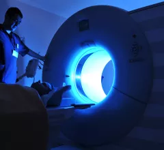
Magnetic resonance imaging (MRI) is the gold standard imaging modality for soft tissues. It produces detail cross-sectional images of soft tissue and bone anatomy, including muscles, tendons, ligaments, brain and organs, without the use of ionizing radiation. In addition to orthopedic imaging, MRI is also used for heart, brain and breast. MRI uses gadolinium contrast in many exams to highlight tissues and blood vessels, which enhances images and offers better diagnostic quality. It can also be used in conjunction with PET scans. How does MRI work? MR creates images by using powerful magnets to polarize hydrogen atoms in water (the body is made of of more than 80% water) so they face in one direction. A radiofrequency pulse is then used to ping these atoms, causing them to wobble, or resonate. The MRI coils detect this and computers can assemble images from the signals. Basic MRI scans will focus on the resonance of fat and water in two different sequences, which highlight and contrast different features in the anatomy.

Thumbnail

‘Quantum leap’: Researchers develop ‘intelligent’ new material for quicker, cheaper MRIs

If such technology is made available commercially, it could revolutionize magnetic resonance imaging, noted Stephan Anderson, a Boston Medical Center radiologist and BU School of Medicine professor of radiology. 

Thumbnail

FDA approves GE Healthcare’s MRI contrast agent for US use

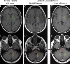
Clariscan is a gadolinium-based agent indicated for IV use in brain, spine and associated tissues to help detect areas with disruption of the blood brain barrier, or abnormal vascularity.

Thumbnail

FDA approves first AI-assisted cardiac MRI scanner

Los Altos, California-based HeartVista has gained approval from the Food and Drug Administration for its one-click heart MRI examination tool. 

Thumbnail

MRI technique may make diagnosing liver cancer easier, scientists say

Experts with Charité – Universitätsmedizin Berlin have developed a new diagnostic technique to let doctors visualize liver tumors using “tomoelastography,” which combines tomography and elasticity.

Thumbnail

AI can predict MR sequence types, saving providers time

Researchers found that deep convolutional neural networks (CNNs) can predict sequence types for brain MR images, sharing their findings in the Journal of Digital Imaging.

Thumbnail

Startup collaborating with Yale on new point-of-care MRI system

As part of the collaboration, Yale New Haven Hospital has been using the MRI system for a two-year study involving the American Heart Association. Results of the preliminary research will be announced early next year.

Thumbnail

Yale School of Medicine, tech company collaborate to create portable MRI

The Ivy League institution is now trying out the transportable tool in the neuro intensive care unit of Yale New Haven Hospital.

Thumbnail

Children’s National Hospital using sophisticated MRI to track impact of opioids on infant brains

The District of Columbia-based provider joins several other institutions as part of the NIH’s massive Helping to End Addiction Long-Term Initiative. 

Around the web

The nuclear imaging isotope shortage of molybdenum-99 may be over now that the sidelined reactor is restarting. ASNC's president says PET and new SPECT technologies helped cardiac imaging labs better weather the storm.

CMS has more than doubled the CCTA payment rate from $175 to $357.13. The move, expected to have a significant impact on the utilization of cardiac CT, received immediate praise from imaging specialists.

The all-in-one Omni Legend PET/CT scanner is now being manufactured in a new production facility in Waukesha, Wisconsin.