Magnetic Resonance Imaging

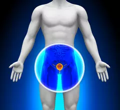
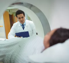
Magnetic resonance imaging (MRI) is the gold standard imaging modality for soft tissues. It produces detail cross-sectional images of soft tissue and bone anatomy, including muscles, tendons, ligaments, brain and organs, without the use of ionizing radiation. In addition to orthopedic imaging, MRI is also used for heart, brain and breast. MRI uses gadolinium contrast in many exams to highlight tissues and blood vessels, which enhances images and offers better diagnostic quality. It can also be used in conjunction with PET scans. How does MRI work? MR creates images by using powerful magnets to polarize hydrogen atoms in water (the body is made of of more than 80% water) so they face in one direction. A radiofrequency pulse is then used to ping these atoms, causing them to wobble, or resonate. The MRI coils detect this and computers can assemble images from the signals. Basic MRI scans will focus on the resonance of fat and water in two different sequences, which highlight and contrast different features in the anatomy.

Thumbnail

ACR publishes new guidance on prostate MRI utilization

The American College of Radiology (ACR) has developed a new guidance document on the use of prostate MRI for working up suspected or known malignancies.

Thumbnail

AI techniques detect implantable devices, improve MRI safety

Two different AI-based methods can identify patients with implantable devices that could pose a safety risk during MRI scans, according to research published in the Journal of the American College of Radiology.

FDA approves use of Boston Scientific DBS system during full-body MRI scans

Boston Scientific Corporation announced that it has received FDA approval for its Vercise Gevia deep brain stimulation (DBS) system to be used during full-body MRI scans.

Thumbnail

Gadolinium-free contrast agent could improve MRI safety

A new manganese-based contrast agent could help limit the use of gadolinium-based contrast agents (GBCAs) and make MRI scans safer, according to findings published in Investigative Radiology.

MRI tech company Metrasens files patent infringement case

Metrasens, a Malvern, England-based MRI technology company, has filed a new patent infringement case against Nanjing Cloud Magnet Electronic Technology Limited (Cloud Magnet).

Thumbnail

MRI scans reveal 'big step' for progressive MS research

Chronic active lesions visible on brain MRI scans, identifiable by their “darkened outer rims,” are associated with multiple sclerosis, according to new findings published in JAMA Neurology.

Thumbnail

How MRI scans can dramatically improve prostate cancer diagnosis

Multiparametric MRI (mpMRI) scans and targeted biopsies can improve the detection of clinically significant prostate cancer, according to new research published in JAMA Network Open.

Thumbnail

FDA shares new guidance document on MRI safety

The FDA has published a new guidance document focused on making improvements to the overall safety of MR environments and MRI-related medical device labeling.

Around the web

The nuclear imaging isotope shortage of molybdenum-99 may be over now that the sidelined reactor is restarting. ASNC's president says PET and new SPECT technologies helped cardiac imaging labs better weather the storm.

CMS has more than doubled the CCTA payment rate from $175 to $357.13. The move, expected to have a significant impact on the utilization of cardiac CT, received immediate praise from imaging specialists.

The all-in-one Omni Legend PET/CT scanner is now being manufactured in a new production facility in Waukesha, Wisconsin.