Magnetic Resonance Imaging

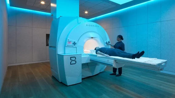
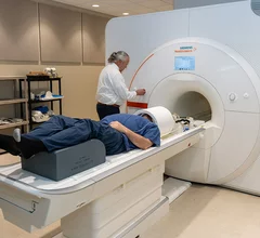
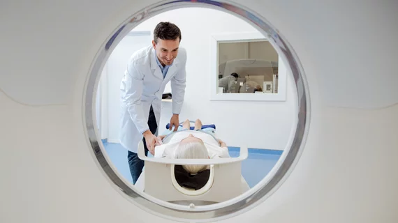
Magnetic resonance imaging (MRI) is the gold standard imaging modality for soft tissues. It produces detail cross-sectional images of soft tissue and bone anatomy, including muscles, tendons, ligaments, brain and organs, without the use of ionizing radiation. In addition to orthopedic imaging, MRI is also used for heart, brain and breast. MRI uses gadolinium contrast in many exams to highlight tissues and blood vessels, which enhances images and offers better diagnostic quality. It can also be used in conjunction with PET scans. How does MRI work? MR creates images by using powerful magnets to polarize hydrogen atoms in water (the body is made of of more than 80% water) so they face in one direction. A radiofrequency pulse is then used to ping these atoms, causing them to wobble, or resonate. The MRI coils detect this and computers can assemble images from the signals. Basic MRI scans will focus on the resonance of fat and water in two different sequences, which highlight and contrast different features in the anatomy.
Thumbnail

A significant amount of imaging orders in emergency settings are inappropriate

A general lack of awareness pertaining to ACR appropriateness criteria could be a driving factor behind the misguided requisitions, authors of a new analysis suggest. 

Alzheimer’s imaging experts to assess utility, efficiency of Hyperfine’s Swoop system

Is portable MRI suitable for finding abnormalities in the brains of patients receiving new amyloid-targeting therapy for Alzheimer’s disease? Clinical researchers are about to find out.

Kim Tzoumakas

Private equity-backed radiology provider Rayus partners with AI startup to launch nationwide whole-body MRI offering

Ezra charges $950 to $2,500 for exams of healthy individuals and analyzes them with its FDA-cleared software to diagnose cancer and other conditions.  

Medicare drops AUC requirement for advanced imaging, ASNC celebrates

"This was an unneeded burden, which was solely adding to the administrative hassles of medicine," said American Society of Nuclear Cardiology President Larry Phillips.

Auburn University SIemens 7T magnetom scanner

University charts a world’s first with clinical use of new $9M 7T MRI system

The Siemens Magnetom scanner utilizes dedicated radiofrequency sodium coils and parallel transmit technology—a “stark” difference from older models

Thumbnail

Deep learning-based MRI reconstruction software produces considerable cost savings

Oulu University Hospital believes AI will allow it to deliver the same level of service with one fewer scanner, enabling annual savings of over $436,000.

Radiologist using the Philips Smart Quant 3D Neuro artificial intelligence (AI) software to perform our measurements for white matter, gray matter and other parameters on brain MRI.

PHOTO GALLERY of brain imaging

This is a clinical photo gallery of neuro imaging and what conditions can be can be visualized in brain scans, and various imaging techniques used.

Medical 3D printing expert Summer Decker, PhD, professor and vice chair for research and innovation, and director of 3D clinical applications, University of South Florida (USF) Morsani College of Medicine and Tampa General Hospital, explains what is involved in creating a 3D printiung program in radiology and how it helps patient care.

Pitching the business case for 3D printing labs in radiology

Medical 3D printing expert Summer Decker, PhD, University of South Florida, explains how her lab has helped improve patient care. 

Around the web

The ACR hopes these changes, including the addition of diagnostic performance feedback, will help reduce the number of patients with incidental nodules lost to follow-up each year.

And it can do so with almost 100% accuracy as a first reader, according to a new large-scale analysis.

The patient, who was being cared for in the ICU, was not accompanied or monitored by nursing staff during his exam, despite being sedated.