Magnetic Resonance Imaging

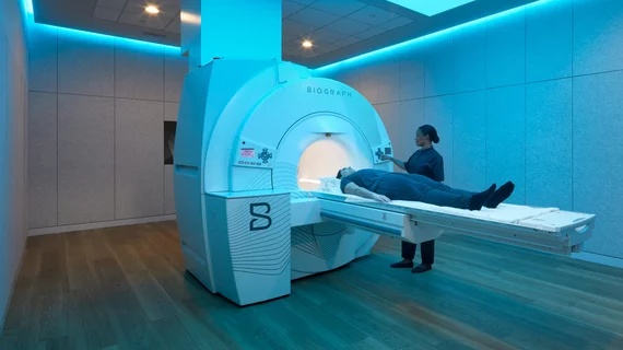
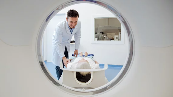
Magnetic resonance imaging (MRI) is the gold standard imaging modality for soft tissues. It produces detail cross-sectional images of soft tissue and bone anatomy, including muscles, tendons, ligaments, brain and organs, without the use of ionizing radiation. In addition to orthopedic imaging, MRI is also used for heart, brain and breast. MRI uses gadolinium contrast in many exams to highlight tissues and blood vessels, which enhances images and offers better diagnostic quality. It can also be used in conjunction with PET scans. How does MRI work? MR creates images by using powerful magnets to polarize hydrogen atoms in water (the body is made of of more than 80% water) so they face in one direction. A radiofrequency pulse is then used to ping these atoms, causing them to wobble, or resonate. The MRI coils detect this and computers can assemble images from the signals. Basic MRI scans will focus on the resonance of fat and water in two different sequences, which highlight and contrast different features in the anatomy.
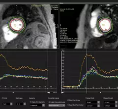
An example of cardiac MRI myocardial blood flow reserve (MBFR) imaging. Image courtesy of https://doi.org/10.3390/diagnostics10090679

Imaging group thrilled with new CPT codes for MBFR measurements

The Society for Cardiovascular Magnetic Resonance applauded two new CPT Category III codes for myocardial blood flow reserve (MBFR) assessments.

Price shopping transparency

‘MRI’ is the most-searched medical term among patients seeking cost information

“CT scan” was the third most-searched term among healthcare consumers, and “ultrasound” was No. 4, according to Fair Health. 

profits revenue finance money

Radiology practice SimonMed doubling centers offering full-body MRI scans amid ‘exponential’ demand growth

The private equity-backed provider first launched SimonOne nationwide in summer 2023, capitalizing on increased interest from startup firms and celebrities. 

Ezra

Whole-body MRI startup Ezra raises $21M, eyes 50 new locations in 2024

Supported by AI, Ezra offers a 60-minute, full-body MRI exam for $1,950, allowing patients to seek out health concerns before they surface. 

Accelerated MRI sequence helps radiologists assess heart disease patients without breath-holding

French imaging experts utilized an investigational, free-breathing, short-axis imaging sequence that deployed deep learning to reconstruct images. 

Medical malpractice gavel diagnostic error mistake stethoscope

Patient sues SimonMed Imaging claiming tech injected her with Valium instead of contrast media prior to MRI

Plaintiff attorneys also allege the sedative was administered in an excessive dose, forcing the woman to go back on anti-seizure meds and delay her fertility treatment. 

doors subspecialty specialize

Inconsistent imaging: Radiologists letting personal preferences influence decision-making

There is significant variation from one radiologist to the next in suggesting follow-up head and neck imaging, Harvard experts detailed in AJR

SimonMed

Private equity-backed radiology provider SimonMed opens 6 new imaging centers

New additions balloon the Scottsdale, Arizona-based radiology practice's total number of locations to 170, spanning 11 states. 

Around the web

The ACR hopes these changes, including the addition of diagnostic performance feedback, will help reduce the number of patients with incidental nodules lost to follow-up each year.

And it can do so with almost 100% accuracy as a first reader, according to a new large-scale analysis.

The patient, who was being cared for in the ICU, was not accompanied or monitored by nursing staff during his exam, despite being sedated.