
Ultrasound is both the prevention and solution for cosmetic filler-related injuries
New data shared this week during RSNA suggests that ultrasound is key for both the prevention and treatment of adverse events related to the injection of cosmetic fillers.
Facial fillers have become increasingly popular in recent years, with hyaluronic acid-based fillers being the most prevalent. Although these injections are fairly routine, they are not without risks.
When fillers are injected without imaging guidance, there is the risk that the materials could be misplaced. A common complication of this scenario is vascular occlusion, which occurs when filler is injected in a way that disrupts bloodflow to arteries. The area around the nose—a popular location for filler injections—is especially vulnerable to this complication due to the surrounding blood vessels that communicate with the external and internal carotid system. Occlusion in this area could lead to serious complications, such as blindness, stroke and more experts involved in the research caution.
“Vascular occlusion events in the face can be devastating, because, if they’re not properly treated, they can cause necrosis and even facial deformation,” Rosa Maria Silveira Sigrist, MD, an attending radiologist and PhD candidate at the University of São Paulo Department of Radiology in Brazil, and co-authors explain.
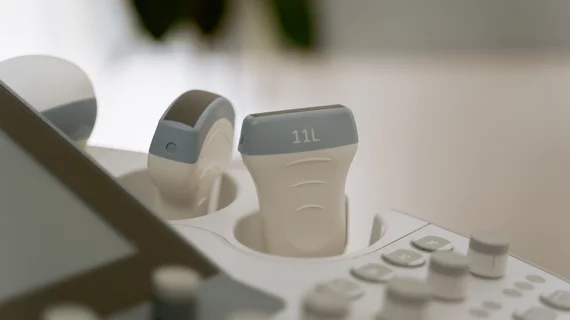
Sigrist and colleagues recently evaluated 100 cases of filler-related vascular complications that occurred over a span of 3 years across 4 radiology centers, 1 dermatology center and 1 plastic surgery center. Each case involved ultrasound imaging to help providers assess vascular injuries.
Imaging revealed decreased or absent flow to the perforator vessels, which connect superficially to deep arteries in the face, in 42% of cases. Concerningly, in 35% of these instances, bloodflow had been occluded from the lateral nasal arteries, putting patients at serious risk.
In cases of misplaced filler, a substance called hyaluronidase can be injected to break down the hyaluronic acid and relieve the occlusions. However, these injections should be guided by ultrasound to ensure proper placement and reduce the risk of additional vascular injury.
“If injectors are not guided by ultrasound, they treat based on where the clinical findings are and inject blindly,” Sigrist notes. “But if we can see the ultrasound finding, we can target the exact place where the occlusion occurs. Rather than flooding the area with hyaluronidase, we can do guided injections that use less hyaluronidase and provide better treatment results.”
Sigrist went on to suggest that ultrasound could prevent these complications from occurring in the first place, further supporting the use of imaging in cosmetic settings.

In addition to her background in journalism, Hannah also has patient-facing experience in clinical settings, having spent more than 12 years working as a registered rad tech. She began covering the medical imaging industry for Innovate Healthcare in 2021.