
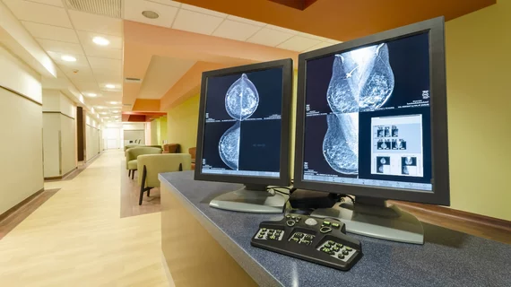
The BI-RADS 2025 Manual is now live—here are a few of its key updates
The Breast Imaging Reporting and Data System (BI-RADS) 2025 Manual was unveiled Monday during an educational session at this year’s annual meeting of the Radiological Society of North America.
Several experts who contributed to the manual’s updates spoke on the specifics of what has changed and how these revisions will affect providers. Speakers touched on each of the breast imaging modalities, highlighting details pertinent to everyday practice.
"The BI-RADS Manual is an invaluable resource for breast imagers to clearly communicate breast imaging findings in everyday practice,” Mary S. Newell, MD, chair of the ACR Committee on BI-RADS, said in a statement. “The updates to this manual, which is the culmination of several years of work from the ACR BI-RADS committee and subcommittee members, will assist with the peer review process when auditing breast imaging practices, which is important for identifying opportunities for practice improvement.”
The committee sought to provide a manual that supported structured, modality-neutral reporting that would help readers more accurately describe findings that could impact patient management.
Here are some of the highlights:
Imaging examples have been updated to include digital views.
New search functions were developed to optimize provider queries on specific scenarios.
A guidance chapter for digital breast tomosynthesis was added. The goal of this chapter is to aid interpreting radiologists with the management of certain clinical scenarios and findings, and to improve consistency of DBT reporting.
The DBT section also includes an update to how lesions assessed on only one view can be described. Previously, masses seen on only one view could not be referred to as lesions. The update now allows these findings to be described as lesions; it is the committee’s hope this could eliminate the need for additional mammography views and radiation exposure.
Breast density assessments have been updated to reflect the likelihood overlying tissue may obscure cancer. Previously, such assessments were based on a percentage of dense tissue. Breast density classification is now based on the densest region, not the entire breast. The group also added a subsection on risk significance relative to breast density.
New mass descriptors have been added and removed for all modalities. They pertain to size, shape, associated features and margins.
Lymph nodes now have their own dedicated section for each modality (they were previously included in “associated features”).
New information and recommendations for implants and gynecomastia have been added, as has guidance on special cases, such as mastectomies and abscesses.
A new section on contrast-enhanced mammography has been added. This suggests that providers should describe low energy images separately from recombined one. The low energy portion will use the same lexicon as mammography, while the recombined images will use terms adopted from the MRI section but modified to cover situations unique to CEM.
Updates to tissue composition for breast ultrasound have been added.
Ultrasound revisions also include information on non-mass lesions for clarification.
Breast MRI updates describe different acquisition parameters (fast protocols, DWI, etc.) and subsequent findings for each.
The breast MRI lexicon has added a Mass T2 Hyperintensity Descriptor.
Additional guidance on the management of categories 3 and 6 (for MRI) has been added to provide clarification on how to manage specific patient scenarios.
The full updated manual went live today. It can be found on the American College of Radiology’s website here.

In addition to her background in journalism, Hannah also has patient-facing experience in clinical settings, having spent more than 12 years working as a registered rad tech. She began covering the medical imaging industry for Innovate Healthcare in 2021.