Computed Tomography

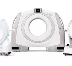
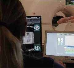
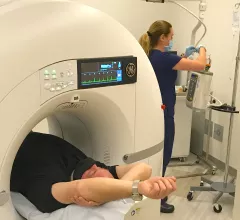
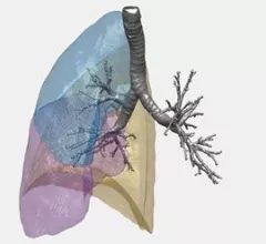
Computed tomography (CT) is a fast and accurate imaging modality often used in emergency settings and trauma imaging. CT scans, with or without (or both) iodinated contrast are frequently used to image the brain, chest, abdomen and pelvis, but also have post-imaging reconstructive capabilities for detailed orthopedic imaging. It is now a standard imaging modality in emergency rooms to quickly assess patients. CT uses a series of X-ray images shot as the gantry rotates around the patient. Computer technology assembles these into into a dataset volume than can be slices on any access, or advanced visualization software can extract specific parts of the anatomy for study. Find more content specific to cardiac CT.


VIDEO: Cardiac CT as a front-line chest pain exam in the ED

Harold Litt, MD, explains the use of cardiac CT as a primary imaging exam for chest pain at his emergency department over the past decade.

Iodine contrast being loaded into a contrast injector in preparation for a cardiac CT scan at Duly Health and Care in Lisle, Illinois. The contrast shortage is causing some healthcare organizations to postpone exams and procedures and ration contrast supplies. Photo by Dave Fornell

‘Relatively new’ contrast vendor releases second generic agent

A global tech and pharma supplier that introduced a generic equivalent to GE Healthcare’s Visipaque during the height of the 2022 contrast shortage has begun offering a substitute for Guerbet’s Dotarem.

Thumbnail

Transplanted lungs react to COVID in a distinctive way

Clinicians treating COVID-19 patients who have transplanted lungs and lower airway infection should order molecular testing in addition to, or regardless of, imaging findings.

Long and longer: Imaging wait times in Canada

Prior to the arrival of COVID-19 in early 2020, Canadian patients waited nearly three months for an MRI and more than 11 weeks for a CT. Things have only gotten worse since then.

AI for assessing bone fragility gets breakthrough tag

Orthopedic cone-beam CT supplier CurveBeam AI has received the FDA’s breakthrough device designation for software that computes risk of fracture in patients with osteopenia.

At the site of the ‘God particle,’ explorers advance along a new frontier in medical imaging

The event was the sixth Workshop on Medical Applications of Spectroscopic X-ray Detectors, which wrapped Sept. 1 at the largest particle physics lab in the world.

Viz.ai’s automated ventricle assessor cleared

The FDA has OK’d an algorithm that automatically ratios the diameter of the right ventricle vs. that of the left on CT pulmonary angiography studies for patients with pulmonary embolism (PE).

Ischemic stroke shown in CT scans. Image courtesy of RSNA

VIDEO: AI for stroke detection on CT imaging

Bibb Allen, MD, FACR, chief medical officer of the American College of Radiology (ACR) Data Science Institute, explains the trend of using AI for the automated detection of stroke on computed tomography (CT) imaging and the need to include radiologists on the stroke care team.

Around the web

The all-in-one Omni Legend PET/CT scanner is now being manufactured in a new production facility in Waukesha, Wisconsin.

After reviewing years of data from its clinic, one institution discovered that issues with implant data integrity frequently put patients at risk. 

Prior to the final proposal’s release, the American College of Radiology reached out to CMS to offer its recommendations on payment rates for five out of the six the new codes.

Trimed Popup
Trimed Popup