Computed Tomography

Computed tomography (CT) is a fast and accurate imaging modality often used in emergency settings and trauma imaging. CT scans, with or without (or both) iodinated contrast are frequently used to image the brain, chest, abdomen and pelvis, but also have post-imaging reconstructive capabilities for detailed orthopedic imaging. It is now a standard imaging modality in emergency rooms to quickly assess patients. CT uses a series of X-ray images shot as the gantry rotates around the patient. Computer technology assembles these into into a dataset volume than can be slices on any access, or advanced visualization software can extract specific parts of the anatomy for study. Find more content specific to cardiac CT.


Thumbnail

Emergency trunk CT rises sharply even for minor, moderate trauma

Almost 10 of every 1,000 patients evaluated for trauma injuries in 2018 received CT scans of the chest, abdomen and pelvis to supply a diagnostic view of, in essence, the entire torso.

Thumbnail

5 imaging modalities have changed the war on cancer over the past 50 years. How many can you name?

Two branch chiefs with the cancer imaging program at NIH’s National Cancer Institute have produced a written commemoration to mark the golden anniversary of 1971’s National Cancer Act.

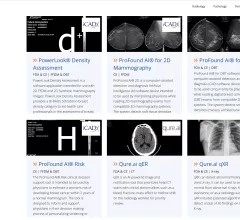
Example of an artificial intelligence (AI) app store on the Sectra website, where Sectra PACS users can select the AI algorithms they want that are already integrated into the Sectra System. Other vendors have followed a similar approach to AI developed by many smaller vendors they partner with.

VIDEO: Development of AI app stores to enable easier access

Keith J. Dreyer, DO, PhD, FACR, American College of Radiology (ACR) Data Science Institute Chief Science Officer, explains how radiology vendors have developed AI app stores to make it easier to access new FDA cleared AI algorithms.
 

Keith J. Dreyer, DO, PhD, FACR, American College of Radiology (ACR) Data Science Institute Chief Science Officer, explains artificial intelligence (AI) for radiology. Dreyer also holds the positions of vice chairman of radiology at Massachusetts General Hospital, chief data science and information officer for the departments of radiology for both Massachusetts General Hospital and Brigham and Women's Hospital.

VIDEO: Where will radiology AI be in 5 years?

Keith J. Dreyer, DO, PhD, FACR, American College of Radiology (ACR) Data Science Institute Chief Science Officer, explains 5 developments to watch for in radiology artificial intelligence (AI).

Emergency overreads change care for 92% of patients with discrepant outside reports

When emergency radiologists overread interpretations of critically ill transfer patients, they discover discrepancies in fewer than 15% of cases. However, more than 90% of these second opinions produce a change in patient care or follow-up.

Faster CT translates to fewer sedated children

Upgrading to dual-source, dual-energy CT machines cut average pediatric scan times from around 12 seconds to three seconds or less at two sites of an academic emergency department.

FDA greenlights ortho robot, automated MRI brain modeling

FDA approvals arrive for ClearPoint Neuro and MicroPort Navibot. 

COVID-19 medical imaging examples of various clinical presentations. SARS-CoV-2 clinical imaging presentations.

PHOTO GALLERY: What does COVID-19 look like on medical imaging?

This image gallery shows what the various clinical presentations associated with the COVID-19 virus that have been documented during the coronavirus pandemic.

Around the web

The all-in-one Omni Legend PET/CT scanner is now being manufactured in a new production facility in Waukesha, Wisconsin.

After reviewing years of data from its clinic, one institution discovered that issues with implant data integrity frequently put patients at risk. 

Prior to the final proposal’s release, the American College of Radiology reached out to CMS to offer its recommendations on payment rates for five out of the six the new codes.

Trimed Popup
Trimed Popup